. 2021 Aug 17;10(8):2108.
doi: 10.3390/cells10082108.

Unraveling Human AQP5-PIP Molecular Interaction and Effect on AQP5 Salivary Glands Localization in SS Patients

Affiliations

Unraveling Human AQP5-PIP Molecular Interaction and Effect on AQP5 Salivary Glands Localization in SS Patients

Clara Chivasso et al. Cells. .

Abstract

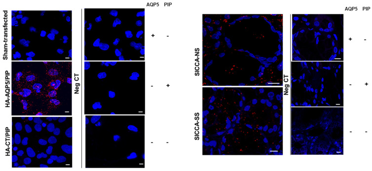
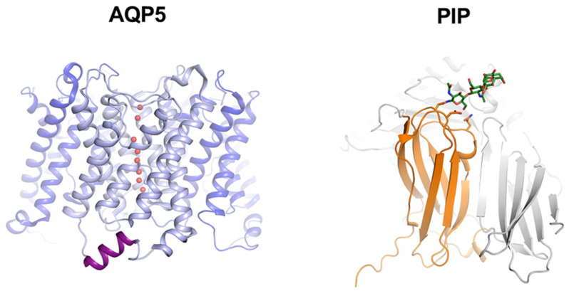
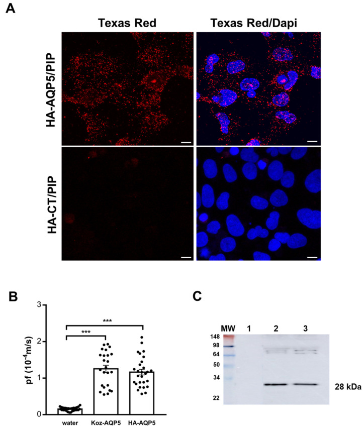
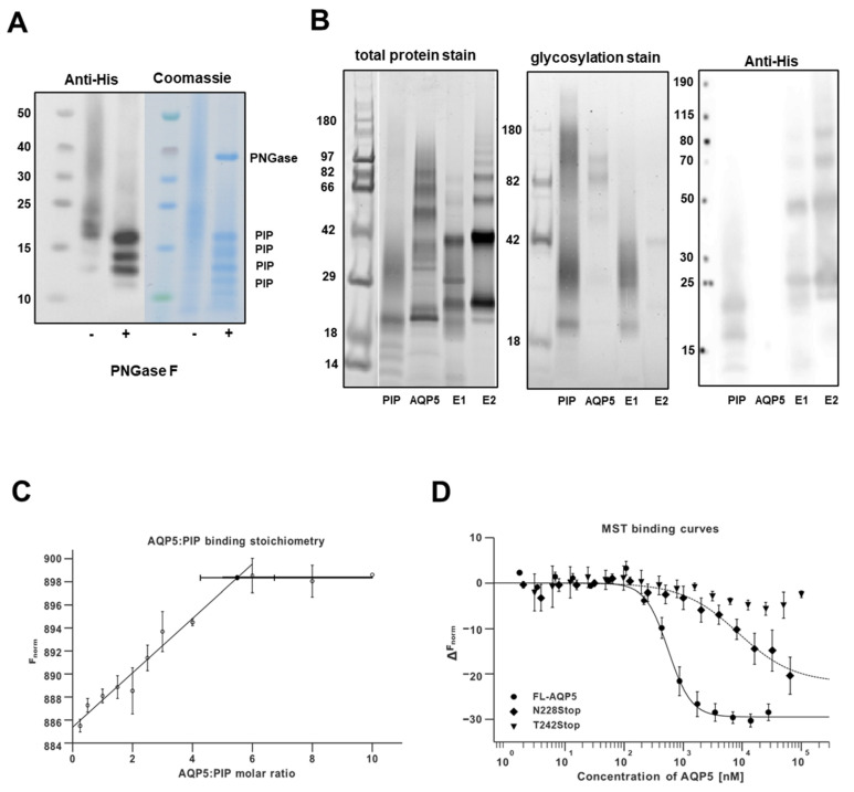
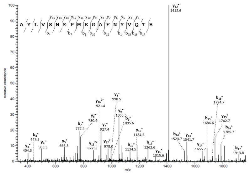
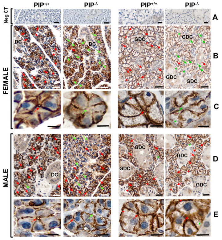
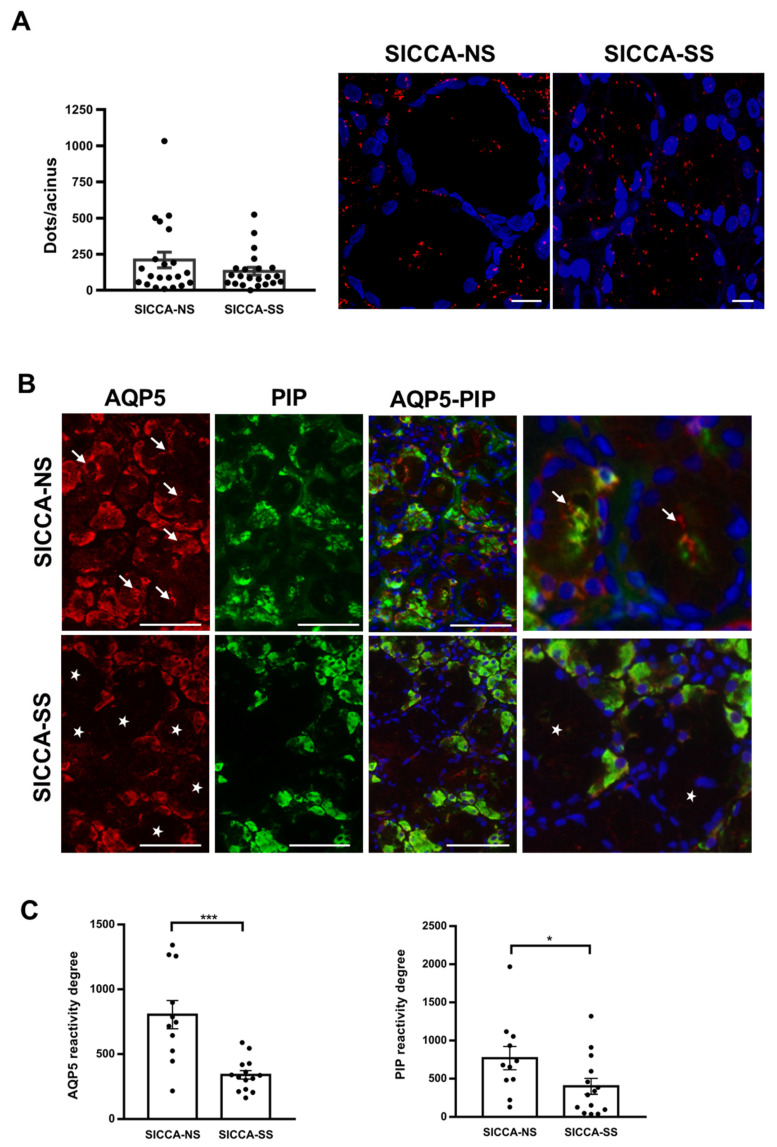
Saliva secretion requires effective translocation of aquaporin 5 (AQP5) water channel to the salivary glands (SGs) acinar apical membrane. Patients with Sjögren's syndrome (SS) display abnormal AQP5 localization within acinar cells from SGs that correlate with sicca manifestation and glands hypofunction. Several proteins such as Prolactin-inducible protein (PIP) may regulate AQP5 trafficking as observed in lacrimal glands from mice. However, the role of the AQP5-PIP complex remains poorly understood. In the present study, we show that PIP interacts with AQP5 in vitro and in mice as well as in human SGs and that PIP misexpression correlates with an altered AQP5 distribution at the acinar apical membrane in PIP knockout mice and SS hMSG. Furthermore, our data show that the protein-protein interaction involves the AQP5 C-terminus and the N-terminal of PIP (one molecule of PIP per AQP5 tetramer). In conclusion, our findings highlight for the first time the role of PIP as a protein controlling AQP5 localization in human salivary glands but extend beyond due to the PIP-AQP5 interaction described in lung and breast cancers.

Keywords: Sjögren’s syndrome; aquaporin-5; prolactin-inducible protein; salivary gland.


Conflict of interest statement

The authors declare no conflict of interest.

Figures

Figure A1
Figure A1
Protein-protein interactions in transfected NS-SV-AC cells and hMSG. Protein-protein interactions visualized as red fluorescent spots on transfected NS-SV-AC (left) and hMSG (right). Nuclei were labelled with DAPI (blue). Left, negative controls were performed using NS-SV-AC transfected with HA-AQP5 and PIP in the absence of anti-HA antibody, anti-PIP or both primary antibodies. Right, negative controls were performed using hMSG from SICCA-NS in the absence of anti-AQP5 antibody, anti-PIP or both primary antibodies. Scale bar, 10 µm.
Figure 1
Figure 1
Left: crystal structure of the human AQP5 tetramer (PDB code 3D9S) with one monomer highlighted in light blue and water molecules in red. The C-terminal helix that is known to be involved in protein-protein interactions in other AQPs is highlighted in purple. The last 21 amino acids were not resolved in the crystal structure and are therefore not shown. Right: crystal structure of human PIP (orange) in complex with the Zinc α2-glycoprotein (ZAG, grey) (PDB code 3ES6). A single glycosylation chain on Asn77 in PIP is shown in green.
Figure 2
Figure 2
(A) PLA performed on transfected NS-SV-AC cells with HA-CT and PIP and HA-AQP5 and PIP. Nuclei were labeled with DAPI (blue) and AQP5-PIP interactions are in red (Texas Red). Scale bar, 10µm. Negative controls are shown in Appendix A, Figure A1, left. Images are representative of 3 experiments. (B) Xenopus laevis oocytes swelling assay. Results are the mean ± S.E.M. (n = 29 for water; n = 24 for Koz-AQP5; n = 28 for HA-AQP5). Statistical analysis was performed using One-way ANOVA; ***: p < 0.001. (C) The expression of AQP5 was verified by WB in the injected oocytes.
Figure 3
Figure 3
AQP5 and PIP interact at molecular levels. (A) WB and SDS-PAGE gel of PIP before and after deglycosylation. (B) SDS-PAGE and WB of E1 and E2 samples from the co-elution assay. (C) MST binding curves for PIP binding to AQP5 constructs (n = 3). (D) Stoichiometry analysis of FL-AQP5 binding to PIP (n = 2). Results are expressed as the mean ± S.E.M.
Figure 4
Figure 4
PIP interacts with AQP5 in mouse SGs. Tandem mass spectra of PIP peptides obtained following AQP5 immunoprecipitation in mouse SGs.
Figure 5
Figure 5
AQP5 localization in P and SM in PIP+/+ and PIP−/− male and female mice. Negative control (Neg CT-up panels) was performed in the absence of a primary antibody. Images are representative of 4 PIP+/+ and 4 PIP−/− male mice for P glands, 5 mice for other groups. Red and green arrows indicate respectively the presence or absence of apical staining in acinar cells. Scale bar: 20 µm (images in horizontal lanes (A,B,D)) and 10 µm (images in horizontal lanes (CE)).
Figure 6
Figure 6
Sjögren’s syndrome alters the expression of AQP5-PIP complexes, and AQP5 and PIP in hMSG. ((A) Left), quantification of PLA red spots per acinus composed of 15 acinar cells. Results are expressed as the mean ± S.E.M cells (n = 21 SICCA-NS, 22 SICCA-SS). Statistical analysis was performed using Mann Whitney u test; p > 0.05. ((A) Right), PLA on hMSG biopsy from SICCA-NS and SICCA-SS. Nuclei were labeled with DAPI (blue) and interactions are represented by red spots. Scale bar, 10 µm. (B) Immunofluorescence of AQP5 (red) and PIP (green) in SICCA-NS and SICCA-SS hMSG. Scale bar; 75 µm. Negative controls are shown in Appendix A Figure A1, right. (C) Semi-quantitative evaluation of AQP5 and PIP expression. Results are expressed as the mean ± S.E.M. (n = 11 SICCA-NS, n = 14 SICCA-SS). Statistical analysis was performed using Mann Whitney u test, * p < 0.05; *** p < 0.001.

References

    1. Parisis D., Chivasso C., Perret J., Soyfoo M.S., Delporte C. Current State of Knowledge on Primary Sjögren’s Syndrome, an Autoimmune Exocrinopathy. J. Clin. Med. 2020;9:2299. doi: 10.3390/jcm9072299. - DOI - PMC - PubMed
    1. Agre P. Aquaporin Water Channels (Nobel Lecture) Angew. Chem. Int. Ed. Engl. 2004;43:4278–4290. doi: 10.1002/anie.200460804. - DOI - PubMed
    1. Horsefield R., Nordén K., Fellert M., Backmark A., Törnroth-Horsefield S., van Scheltinga A.C.T., Kvassman J., Kjellbom P., Johanson U., Neutze R. High-Resolution x-Ray Structure of Human Aquaporin 5. Proc. Natl. Acad. Sci. USA. 2008;105:13327–13332. doi: 10.1073/pnas.0801466105. - DOI - PMC - PubMed
    1. Hosoi K., Yao C., Hasegawa T., Yoshimura H., Akamatsu T. Dynamics of Salivary Gland AQP5 under Normal and Pathologic Conditions. Int. J. Mol. Sci. 2020;21:1182. doi: 10.3390/ijms21041182. - DOI - PMC - PubMed
    1. Delporte C. Aquaporins in Secretory Glands and Their Role in Sjögren’s Syndrome. Handb. Exp. Pharmacol. 2009:185–201. doi: 10.1007/978-3-540-79885-9_9. - DOI - PubMed

Publication types

LinkOut - more resources