

The Role of Glycosylation in Melanoma Progression
- PMID: 34440905
- PMCID: PMC8393314
- DOI: 10.3390/cells10082136
The Role of Glycosylation in Melanoma Progression
Abstract
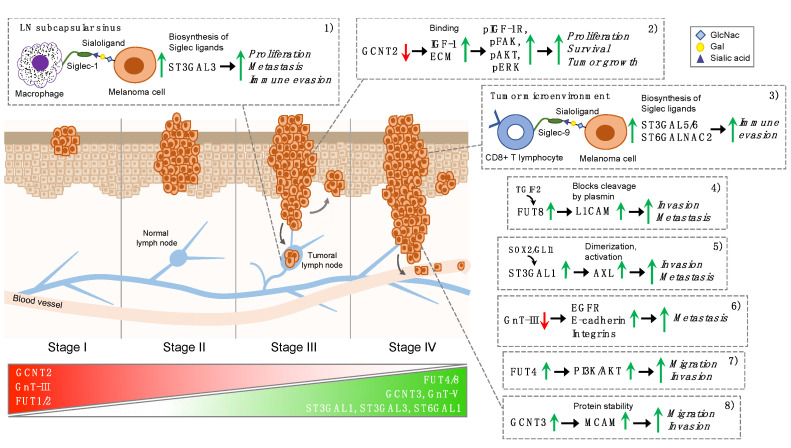
Malignant melanoma is the most aggressive form of skin cancer, which originates from the malignant transformation of melanocytes, the melanin-producing cells of the skin. Melanoma progression is typically described as a stepwise process in which metastasis formation ensues late during disease. A large body of evidence has shown that the accumulation of genetic and epigenetic alterations drives melanoma progression through the different steps. Mortality in melanoma is associated with metastatic disease. Accordingly, early-stage melanoma can be cured in the majority of cases by surgical excision, while late-stage melanoma is a highly lethal disease. Glycosylation is a post-translational modification that involves the transfer of glycosyl moieties to specific amino acid residues of proteins to form glycosidic bonds through the activity of glycosyltransferases. Aberrant glycosylation is considered a hallmark of cancer as it occurs in the majority of tumor types, including melanoma. The most widely occurring glycosylation changes in melanoma are represented by sialylation, fucosylation, and N- and I-glycan branching. In this review, we discuss the role of glycosylation in melanoma and provide insights on the mechanisms by which aberrant glycosylation promotes melanoma progression through activation of invasion and metastasis, immune evasion and cell proliferation.
Keywords: fucosylation; glycan branching; glycosylation; immune evasion; melanoma; metastasis; sialylation.
Conflict of interest statement
The authors declare no conflict of interest.
Figures
Similar articles
-
Glycans in melanoma screening. Part 1. The role of β1,6-branched N-linked oligosaccharides in melanoma.Biochem Soc Trans. 2011 Jan;39(1):370-3. doi: 10.1042/BST0390370. Biochem Soc Trans. 2011. PMID: 21265806
-
Glycosylation in Renal Cell Carcinoma: Mechanisms and Clinical Implications.Cells. 2022 Aug 20;11(16):2598. doi: 10.3390/cells11162598. Cells. 2022. PMID: 36010674 Free PMC article. Review.
-
Glycans in melanoma screening. Part 2. Towards the understanding of integrin N-glycosylation in melanoma.Biochem Soc Trans. 2011 Jan;39(1):374-7. doi: 10.1042/BST0390374. Biochem Soc Trans. 2011. PMID: 21265807
-
Targeting aberrant sialylation and fucosylation in prostate cancer cells using potent metabolic inhibitors.Glycobiology. 2023 Dec 30;33(12):1155-1171. doi: 10.1093/glycob/cwad085. Glycobiology. 2023. PMID: 37847613 Free PMC article.
-
Glycans in melanoma: Drivers of tumour progression but sweet targets to exploit for immunotherapy.Immunology. 2024 Sep;173(1):33-52. doi: 10.1111/imm.13801. Epub 2024 May 14. Immunology. 2024. PMID: 38742251 Review.
Cited by
-
Peak Resembling N-acetylaspartate (NAA) on Magnetic Resonance Spectroscopy of Brain Metastases.Medicina (Kaunas). 2024 Apr 19;60(4):662. doi: 10.3390/medicina60040662. Medicina (Kaunas). 2024. PMID: 38674308 Free PMC article.
-
The melanoma tumor glyco-code impacts human dendritic cells' functionality and dictates clinical outcomes.Front Immunol. 2023 Feb 20;14:1120434. doi: 10.3389/fimmu.2023.1120434. eCollection 2023. Front Immunol. 2023. PMID: 36891308 Free PMC article.
-
Apprising Diagnostic and Prognostic Biomarkers in Cutaneous Melanoma-Persistent Updating.J Pers Med. 2022 Sep 14;12(9):1506. doi: 10.3390/jpm12091506. J Pers Med. 2022. PMID: 36143291 Free PMC article. Review.
-
Role of glycosylation-related gene MGAT1 in pancreatic ductal adenocarcinoma.Front Immunol. 2024 Aug 1;15:1438935. doi: 10.3389/fimmu.2024.1438935. eCollection 2024. Front Immunol. 2024. PMID: 39156890 Free PMC article.
-
A novel glycosyltransferase-related lncRNA signature correlates with lung adenocarcinoma prognosis.Front Oncol. 2022 Aug 18;12:950783. doi: 10.3389/fonc.2022.950783. eCollection 2022. Front Oncol. 2022. PMID: 36059686 Free PMC article.
References
Publication types
MeSH terms
Substances
LinkOut - more resources
Full Text Sources
Other Literature Sources
Medical
