. 2021 Aug 19;57(8):840.
doi: 10.3390/medicina57080840.

Association between Cone-Beam Computed Tomography and Histological and Immunohistochemical Features in Periapical Lesions Correlated with Thickened Maxillary Sinus Mucosa

Affiliations

Association between Cone-Beam Computed Tomography and Histological and Immunohistochemical Features in Periapical Lesions Correlated with Thickened Maxillary Sinus Mucosa

Alexandra Dumitrescu et al. Medicina (Kaunas). .

Abstract

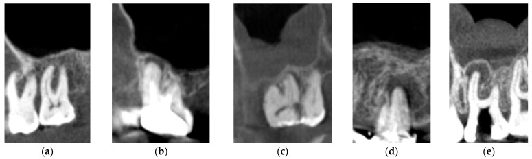
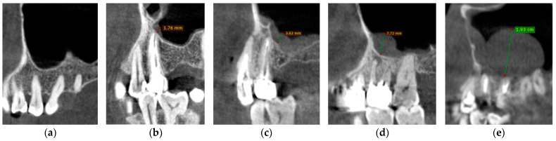
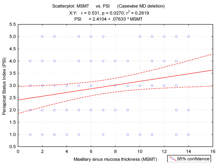
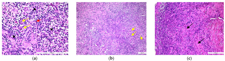
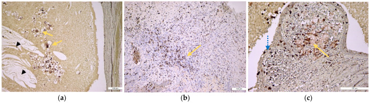
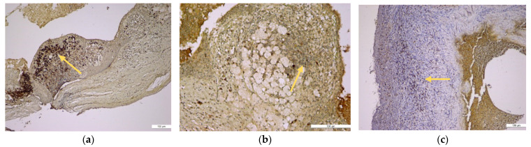
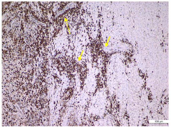
Background and Objectives: Odontogenic sinusitis is a frequently underestimated pathology with fewer symptoms in patients with periapical lesions, periodontal disease, or iatrogenic foreign bodies in the maxillary sinus. The aim of our study was to determine the correlation between maxillary sinusitis and periapical lesions using cone-beam computed tomography (CBCT) imaging and histological and immunohistochemical investigations. Materials and Methods: A total of 1450 initial patients diagnosed with maxillary sinusitis in the Ear-Nose-Throat (ENT) Department, University of Medicine and Pharmacy "Grigore T. Popa" Iasi, Romania, were treated with anti-inflammatory drugs. Of these, 629 still had unresolved symptomatology and were later referred to the Dental Medicine departments for further investigations. Only 50 subjects with periapical lesions in the premolar/molar maxillary area were included in the present study. All the periapical lesions were observed on CBCT and classified using the Periapical Status Index (PSI) and the mean maxillary sinus mucosa thicknesses (MSMT). The enrolled patients underwent surgical procedures with the excision of periapical lesions. The excised samples were submitted to the histological and immunohistochemical investigations. Results: The 50 patients presented periapical lesions of their maxillary teeth in 328 dental units. There was a higher prevalence of periapical lesions in men than in women (chi-square test). We observed a significant difference between the mean MSMT of individuals with periapical lesions compared to those without (p < 0.01). Mean MSMT was 1.23 mm for teeth without periapical lesions and 3.95 mm for teeth with periapical lesions. The histopathological study identified 50% cases with periapical granulomas, 10% cases with periapical granulomas with cystic potential, and 40% cases as periapical cysts. Immunohistochemical stainings showed that CD4+ helper and CD8+ cytotoxic T lymphocytes, along with CD20+ B lymphocytes and CD68+ macrophages, were diffusely distributed in all periapical cysts and in some periapical granulomas, but CD79α+ plasma cells characterized especially periapical granulomas. Conclusions: The current study observed a significant correlation between CBCT maxillary mucosa thickness and type of periapical lesion. Chronic inflammatory lympho-histiocytic infiltrate predominates in periapical lesions, supporting the idea that lesion progression is determined by a humoral-type (CD20+ and CD79α+ B lymphocytes) but also by a cellular-type (CD4+ and CD8+ T lymphocyte population) immune mechanism.

Keywords: cone-beam computed tomography; endo-perio lesions; immunohistochemistry; odontogenic sinusitis; periapical cyst; periapical granuloma.


Conflict of interest statement

The authors declare no conflict of interest.

Figures

Figure 1
Figure 1
CBCT sagittal reconstructions showing the status of periapical region according to the five classes of PSI: (a). Class 1; (b) Class 2; (c) Class 3; (d) Class 4; (e) Class 5.
Figure 2
Figure 2
CBCT sagittal reconstructions showing the maxillary sinus mucosa thickness according to the five classes: (a) Class 1; (b) Class 2; (c) Class 3; (d) Class 4; (e) Class 5.
Figure 3
Figure 3
Correlation between the periapical status index (PSI) and maxillary sinus mucosa thickness. MS, maxillary sinus type.
Figure 4
Figure 4
Histopathological features of periapical granuloma: (a) the early stage revealed a granulation (fibrovascular) tissue infiltrated by a heavy collection of chronic (mononuclear) inflammatory cells (black arrow head) but also few neutrophils (red arrow). There are numerous newly formed small blood vessels (yellow arrow) and thin, immature collagen in the background as well as numerous lymphocytes and plasma cells (HE staining, ×400); (b) the mature stage: granulation (fibrovascular) tissue infiltrated by a collection of chronic (mononuclear) inflammatory cells. There are numerous newly formed small blood vessels and fibrils of collagen in the background as well as numerous lymphocytes and plasma cells but also spindle shape fibroblasts, macrophages (histiocytes), and multinucleated giant cells (yellow arrow) (HE staining, ×100); (c) old stage: large sheets of inflammatory mononuclear cells included into heavy stromal fibrosis (black arrows) (HE staining, ×200).
Figure 5
Figure 5
Histopathological features of periapical cyst: (a) low-power view of a large periapical cyst with a distended lumen that is lined by a non-keratinized stratified squamous epithelium (orange arrow) and is filled with an amorphous eosinophilic material (black arrow head), representing a coagulated fluid high in proteins but also with many cholesterol clefts. The cyst outer wall is thick and is made of fibrous conjunctive tissue (interrupted line arrow) containing a diffuse infiltration of inflammatory mononuclear cells (HE staining, ×10); (b) high-power view of the epithelial lining (orange arrow) but also of the outer fibrous wall that showed a heavy infiltrate with mononuclear inflammatory cells (interrupted line arrow) (HE staining, ×100).
Figure 6
Figure 6
Immunohistochemical identification of the macrophages in the periapical cyst structure. (a) Periapical cyst lumen contained cholesterol crystals (black arrow head) and groups of CD68+ macrophages (orange arrow) (anti-CD68 antibody, ×100); (b) in the conjunctive fibrous tissue, which makes up the outer wall of the periapical cyst, there were numerous CD68+ macrophages (orange arrow) (anti-CD68 antibody, ×100); (c) periapical cyst wall with foamy CD68+ macrophages in the outer fibrous wall (orange arrow) but also into its epithelial lining (blue interrupted arrow) (anti-CD68 antibody, ×200).
Figure 7
Figure 7
Immunohistochemical identification of the lymphocytes in the periapical cyst structure. (a) Numerous B lymphocytes, suggesting a follicular structure (orange arrow) located in the outer fibrous wall (anti-CD20 antibody, ×100); (b) numerous helper T lymphocytes in the outer fibrous wall (orange arrow) (anti-CD4 antibody, ×200); (c) diffuse inflammatory infiltrate made up of cytotoxic T lymphocytes that are located in the outer fibrous wall (orange arrow) (anti-CD8 antibody, ×100).
Figure 8
Figure 8
Immunohistochemical identification of the plasma cells in an old periapical granuloma: large sheets of plasma cells infiltrating the mature granulation tissue (yellow arrow), especially around the blood vessels (interrupted yellow arrow) (anti-CD79α antibody, ×100).

References

    1. Van den Bergh J.P., Ten Bruggenkate C.M., Disch F.J., Tuinzing D.B. Anatomical aspects of sinus floor elevations. Clin. Oral Implants Res. 2000;11:256–265. doi: 10.1034/j.1600-0501.2000.011003256.x. - DOI - PubMed
    1. Lang A.C., Schulze R.K. Detection accuracy of maxillary sinus floor septa in panoramic radiographs using CBCT as gold standard: A multi-observer receiver operating characteristic (ROC) study. Clin. Oral Investig. 2019;23:99–105. doi: 10.1007/s00784-018-2414-1. - DOI - PubMed
    1. Dedeoğlu N., Altun O. Evaluation of maxillary sinus anatomical variations and pathologies in elderly, young, posterior dentate and edentulous patient groups with cone-beam computed tomography. Folia Morphol. 2019;78:595–599. doi: 10.5603/FM.a2019.0013. - DOI - PubMed
    1. Constantine S., Clark B., Kiermeier A., Anderson P. Panoramic radiography is of limited value in the evaluation of maxillary sinus disease. Oral Surg. Oral Med. Oral Pathol. Oral Radiol. Endod. 2019;127:237–246. doi: 10.1016/j.oooo.2018.10.005. - DOI - PubMed
    1. Alsufyani N., El-Hakim H., Major P. Prevalence of maxillary sinus hypoplasia and association with variations in the sinonasal complex: A cone beam CT study. Clin. Oral Investig. 2021;25:5463–5471. doi: 10.1007/s00784-021-03854-3. - DOI - PubMed