Review
. 2021 Jul 26;11(8):1343.
doi: 10.3390/diagnostics11081343.

Laboratory Diagnosis of Porphyria

Affiliations
Review

Laboratory Diagnosis of Porphyria

Elena Di Pierro et al. Diagnostics (Basel). .

Abstract

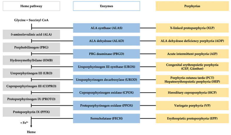
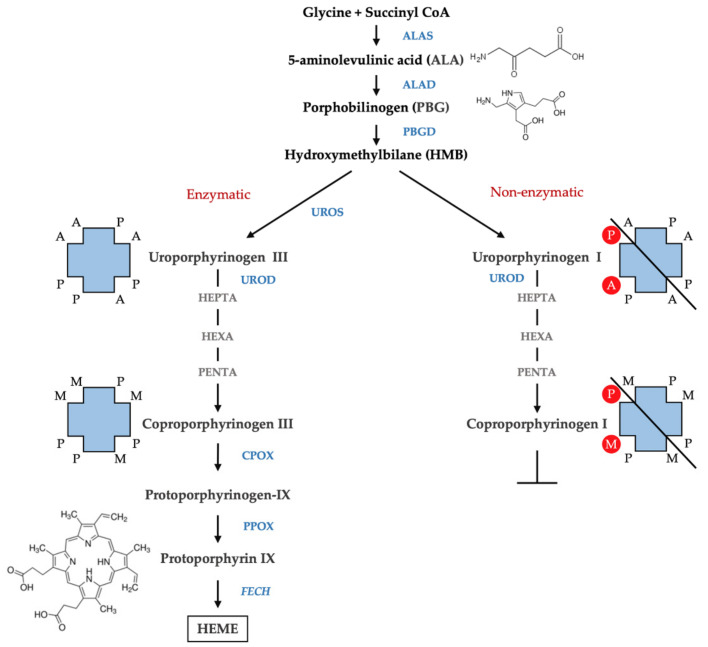
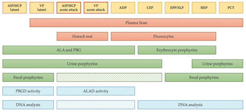
Porphyrias are a group of diseases that are clinically and genetically heterogeneous and originate mostly from inherited dysfunctions of specific enzymes involved in heme biosynthesis. Such dysfunctions result in the excessive production and excretion of the intermediates of the heme biosynthesis pathway in the blood, urine, or feces, and these intermediates are responsible for specific clinical presentations. Porphyrias continue to be underdiagnosed, although laboratory diagnosis based on the measurement of metabolites could be utilized to support clinical suspicion in all symptomatic patients. Moreover, the measurement of enzymatic activities along with a molecular analysis may confirm the diagnosis and are, therefore, crucial for identifying pre-symptomatic carriers. The present review provides an overview of the laboratory assays used most commonly for establishing the diagnosis of porphyria. This would assist the clinicians in prescribing appropriate diagnostic testing and interpreting the testing results.

Keywords: ALA (5-aminolevulinic acid); EPNET (European Porphyria Network); HPLC (high-pressure liquid chromatography); MLPA (multiplex ligation-dependent probe amplification); NGS (next-generation sequencing); PBG (porphobilinogen); diagnosis; porphyria; porphyrins.


Conflict of interest statement

The authors declare no conflict of interest.

Figures

Figure 1
Figure 1
Heme pathway and porphyria. The figure represents the scheme of the heme biosynthesis (in gray), with the involved enzyme (in blue), and the specific porphyria related to each enzyme deficiency (in yellow).
Figure 2
Figure 2
Isomers of porphyrins. The figure represents the scheme of the heme biosynthesis with enzymatic (left) and non-enzymatic conversion of HMB (right). The arrangement of the substituents of the four pyrroles of the porphyrin ring is shown. A, acetate (-CH2COOH); P, propionate (-CH2CH2COOH); M, methyl (-CH3) and vinyl (CH=CH2).
Figure 3
Figure 3
Chromatographic profiles of urine porphyrins. The figure shows the patterns of excretion of healthy individuals (NORMAL) and patients (PCT, HEP, AIP, HCP, VP, CEP, EPP). Fluorescence peaks are identified as uroporphyrin I (1), uroporphyrin III (2), heptacarboxylic acid porphyrin I (3), heptacarboxylic acid porphyrin III (4), hexacarboxylic acid porphyrin III (6), pentacarboxylic acid porphyrin I (7), pentacarboxylic acid porphyrin III (8), coproporphyrin I (10), coproporphyrin III (11).
Figure 4
Figure 4
Chromatographic profiles of fecal porphyrins. The figure shows the patterns of excretion of healthy individuals (NORMAL) and patients (PCT, HEP, AIP, HCP, VP, CEP, EPP). Fluorescence peaks are identified as heptacarboxylic acid porphyrin I (3), heptacarboxylic acid porphyrin III (4), hexacarboxylic acid porphyrin I (5), hexacarboxylic acid porphyrin III (6), pentacarboxylic acid porphyrin I (7), pentacarboxylic acid porphyrin III (8), hydroxyisocoproporphyrin (9), coproporphyrin I (10), coproporphyrin III (11), deethylisocoproporphyrin (12), isocoproporphyrin and dehydroisocoproporphyrin (13), deuteroporphyrin (14), pemptoporphyrin (15), mesoporphyrin (16), protoporphyrin (17).
Figure 5
Figure 5
Laboratory diagnostic strategy. The scheme summarizes the appropriate diagnostic testing to prescribe for each form of porphyria. The striped boxes represent the tests that are not mandatory to prescribe before starting a therapy. In particular, fecal porphyrins and DNA analysis could be avoided during acute attack if other tests are positive and they can be postponed to the phases of latency. Of note, in the sporadic form of PCT, no mutation is expected to be found in UROD gene.

References

    1. Balwani M., Desnick R.J. The porphyrias: Advances in Diagnosis and Treatment. Blood. 2012;120:4496–4504. doi: 10.1182/blood-2012-05-423186. - DOI - PMC - PubMed
    1. Besur S., Hou W., Schmeltzer P., Bonkovsky H.L. Clinically Important Features of Porphyrin and Heme Metabolism and the Porphyrias. Metabolites. 2014;4:977–1006. doi: 10.3390/metabo4040977. - DOI - PMC - PubMed
    1. Stolzel U., Doss M.O., Schuppan D. Clinical Guide and Update on Porphyrias. Gastroenterology. 2019;157:365–381. doi: 10.1053/j.gastro.2019.04.050. - DOI - PubMed
    1. Sassa S. Modern Diagnosis and Management of the Porphyrias. Br. J. Haematol. 2006;135:281–292. doi: 10.1111/j.1365-2141.2006.06289.x. - DOI - PubMed
    1. Wang B., Rudnick S., Cengia B., Bonkovsky H.L. Acute Hepatic Porphyrias: Review and Recent Progress. Hepatol. Commun. 2019;3:193–206. doi: 10.1002/hep4.1297. - DOI - PMC - PubMed

LinkOut - more resources