

Stability of OCT and OCTA in the Intensive Therapy Unit Setting
- PMID: 34441449
- PMCID: PMC8394026
- DOI: 10.3390/diagnostics11081516
Stability of OCT and OCTA in the Intensive Therapy Unit Setting
Abstract
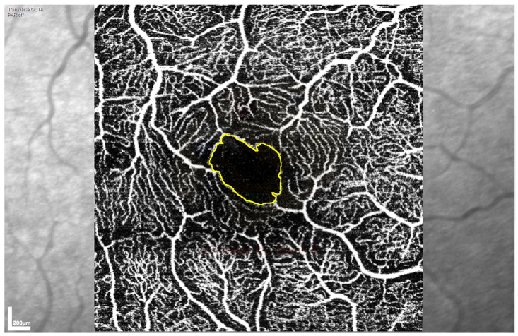
To assess the stability of retinal structure and blood flow measures over time and in different clinical settings using portable optical coherence tomography angiography (OCTA) as a potential biomarker of central perfusion in critical illness, 18 oesophagectomy patients completed retinal structure and blood flow measurements by portable OCT and OCTA in the eye clinic and intensive therapy unit (ITU) across three timepoints: (1) pre-operation in a clinic setting; (2) 24-48 h post-operation during ITU admission; and (3) seven days post-operation, if the patient was still admitted. Blood flow and macular structural measures were stable between the examination settings, with no consistent variation between pre- and post-operation scans, while retinal nerve fibre layer thickness increased in the post-operative scans (+2.31 µm, p = 0.001). Foveal avascular zone (FAZ) measurements were the most stable, with an intraclass correlation coefficient of up to 0.92 for right eye FAZ area. Blood flow and structural measures were lower in left eyes than right eyes. Retinal blood flow assessed in patients before and during an ITU stay using portable OCTA showed no systematic differences between the clinical settings. The stability of retinal blood flow measures suggests the potential for portable OCTA to provide clinically useful measures in ITU patients.
Keywords: critical care; optical coherence tomography angiography; stability.
Conflict of interest statement
The authors declare that they have no competing interests.
Figures
References
-
- Kashani A.H., Chen C.-L., Gahm J.K., Zheng F., Richter G.M., Rosenfeld P.J., Shi Y., Wang R.K. Optical coherence tomography angiography: A comprehensive review of current methods and clinical applications. Prog. Retin. Eye Res. 2017;60:66–100. doi: 10.1016/j.preteyeres.2017.07.002. - DOI - PMC - PubMed
Grants and funding
LinkOut - more resources
Full Text Sources
