Review
. 2021 Aug 17;10(16):3628.
doi: 10.3390/jcm10163628.

Clinical Management of Moyamoya Patients

Affiliations
Review

Clinical Management of Moyamoya Patients

Isabella Canavero et al. J Clin Med. .

Abstract

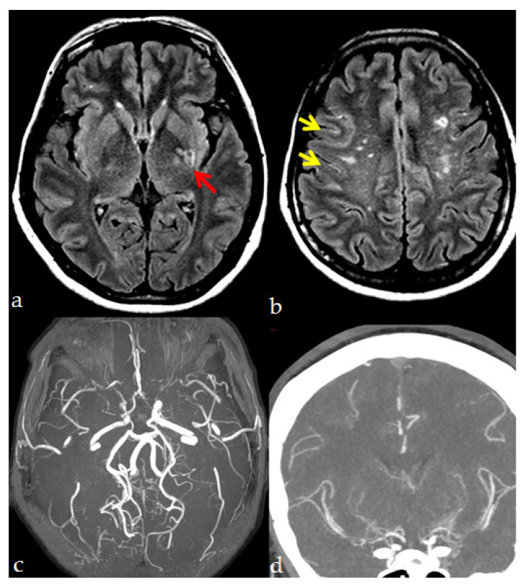
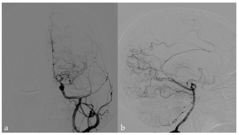
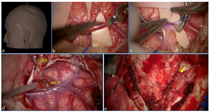
Moyamoya angiopathy (MMA) is a peculiar cerebrovascular condition characterized by progressive steno-occlusion of the terminal part of the internal carotid arteries (ICAs) and their proximal branches, associated with the development of a network of fragile collateral vessels at the base of the brain. The diagnosis is essentially made by radiological angiographic techniques. MMA is often idiopathic (moyamoya disease-MMD); conversely, it can be associated with acquired or hereditary conditions (moyamoya Syndrome-MMS); however, the pathophysiology underlying either MMD or MMS has not been fully elucidated to date, and this poor knowledge reflects uncertainties and heterogeneity in patient management. MMD and MMS also have similar clinical expressions, including, above all, ischemic and hemorrhagic strokes, then headaches, seizures, cognitive impairment, and movement disorders. The available treatment strategies are currently shared between idiopathic MMD and MMS, including pharmacological and surgical stroke prevention treatments and symptomatic drugs. No pharmacological treatment able to reverse the progressive disappearance of the ICAs has been found to date in both idiopathic and syndromic cases. Antithrombotic agents are usually prescribed in ischemic MMA, although the coexisting hemorrhagic risk should be considered. Surgical revascularization techniques, which are currently the best available treatment in symptomatic MMA, are associated with good long-term outcomes and reduced ischemic and hemorrhagic risks. Given the lack of dedicated randomized clinical trials, current treatment is mainly based on observational studies and physicians' and surgeons' expertise.

Keywords: diagnosis; management; moyamoya angiopathy; moyamoya disease; moyamoya syndrome; revascularization; therapy.


Conflict of interest statement

The authors declare no conflict of interest.

Figures

Figure 1
Figure 1
MRI and CT imaging assessment in an MMA patient. Axial FLAIR images show (a) a small lacunar infarction in the left posterior lenticular region, which is pointed out by the red arrow; (b) multiple, non-confluent, white matter hyperintensities with subcortical distribution in both hemispheres; yellow arrows indicate the Ivy sign. (c) MRA with bilateral terminal ICA steno-occlusion with lack of signal in both M1 MCA. (d) CTA in coronal MIP showing the same finding as in (c) with visualization of the distal sylvian segment of MCA and prominent collateralization in the lenticulostriatal perforator vessels. (Imaging performed at the Neuroradiology Department, AUSL Reggio Emilia; pictures are reproduced with patient’s permission).
Figure 2
Figure 2
DSA in an MMA patient (same patient of Figure 1). The left ICA (a) depicts high-grade stenosis of terminal ICA and proximal M1 MCA segment with moyamoya collateralization pattern. Examining the left VA (b), cortical collaterals from PCA and ACA (callosal arcade) to superficial MCA territory are visible. (Imaging performed at the Neuroradiology Department, AUSL Reggio Emilia; pictures are reproduced with patient’s permission).
Figure 3
Figure 3
Direct and indirect revascularization surgery. During surgery, a three-dimensional rendering obtained by CT angiography (a) shows the course of STA (in yellow) and of the peripheral branches of MCA (green) on the left hemisphere in a patient who underwent combined revascularization surgery. The intraoperative pictures acquired under microscopic view depict the different phases of surgical procedure: in (b), temporary clips are positioned in the M4 branch. Then, the STA-MCA bypass is performed with interrupted suture (c). After the bypass is completed (d), the encephaloduromyosynangiosis with dura and temporal muscle (arrow) is then performed (e). (Procedure performed at the Neurosurgery Department, Fondazione IRCCS Istituto Neurologico Carlo Besta, Milan; pictures are reproduced with patient’s permission).

References

    1. Fukui M. Guidelines for the diagnosis and treatment of spontaneous occlusion of the circle of Willis (“moyamoya” disease). Research Committee on Spontaneous Occlusion of the Circle of Willis (Moyamoya Disease) of the Ministry of Health and Welfare, Japan. Clin. Neurol. Neurosurg. 1997;99(Suppl. 2):S238–S240. doi: 10.1016/S0303-8467(97)00082-6. - DOI - PubMed
    1. Scott R.M., Smith E.R. Moyamoya Disease and Moyamoya Syndrome. N. Engl. J. Med. 2009;360:1226–1237. doi: 10.1056/NEJMra0804622. - DOI - PubMed
    1. Guey S., Tournier-Lasserve E., Hervé D., Kossorotoff M. Moyamoya disease and syndromes: From genetics to clinical management. Appl. Clin. Genet. 2015;8:49–68. doi: 10.2147/TACG.S42772. - DOI - PMC - PubMed
    1. Kobayashi E., Saeki N., Oishi H., Hirai S., Yamaura A. Long-term natural history of hemorrhagic moyamoya disease in 42 patients. J. Neurosurg. 2000;93:976–980. doi: 10.3171/jns.2000.93.6.0976. - DOI - PubMed
    1. Bersano A., Guey S., Bedini G., Nava S., Hervé D., Vajkoczy P., Tatlisumak T., Sareela M., Van Der Zwan A., Klijn C.J.M., et al. Research progresses in understanding the pathophysiology of moyamoya disease. Cerebrovasc. Dis. 2016;41:105–118. doi: 10.1159/000442298. - DOI - PubMed