

The Impact of Suture Button Removal in Syndesmosis Fixation
- PMID: 34442022
- PMCID: PMC8397003
- DOI: 10.3390/jcm10163726
The Impact of Suture Button Removal in Syndesmosis Fixation
Abstract
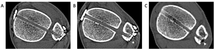
The suture button (SB) device was introduced to negate the need for routine hardware removal in the treatment of syndesmosis injuries. However, a considerable SB removal rate has been reported, and the impact of removal is unknown. This study aimed to evaluate the radiographic and clinical outcomes after removal of SB for syndesmosis fixation. A total of 36 patients who underwent removal surgery after syndesmosis fixation using SB were identified. The mean postoperative time to removal was 12.2 months. On a plain radiograph, tibiofibular clear space (TFCS) was measured and compared at three follow-up time points. In patients with computed tomography (CT) imaging (n = 18), the anterior-to-posterior (A/P) ratio was measured to evaluate changes in quality of reduction. Additionally, clinical outcomes were assessed. There were no significant differences in TFCS between the three follow-up periods. None of the patients exhibited recurrent diastasis after SB removal. Although CT analysis demonstrated malreduction in six patients (33.3%), five of six patients had a subsequent spontaneous reduction of the syndesmosis. Clinically, all patients described the resolution of symptoms related to painful hardware at the final follow-up. Our results demonstrate that SB removal at one year following syndesmosis fixation leads to improved clinical symptoms without negatively impacting the quality of syndesmosis reduction.
Keywords: ankle fracture; diastasis; removal; suture button; syndesmosis; syndesmosis injury; tibiofibular joint.
Conflict of interest statement
None of the authors of this paper has a financial or personal relationship with other people or organizations that could inappropriately influence or bias the content of the paper.
Figures


Similar articles
-
Injury mechanism affects the stability of suture-button syndesmosis fixation.J Orthop Surg Res. 2020 Dec 10;15(1):599. doi: 10.1186/s13018-020-02141-3. J Orthop Surg Res. 2020. PMID: 33302992 Free PMC article.
-
Radiographic Change of the Distal Tibiofibular Joint Following Removal of Transfixing Screw Fixation.Foot Ankle Int. 2018 Mar;39(3):318-325. doi: 10.1177/1071100717745526. Epub 2017 Dec 26. Foot Ankle Int. 2018. PMID: 29278930
-
Maintenance of reduction with suture button fixation devices for ankle syndesmosis repair.Foot Ankle Int. 2015 Jun;36(6):679-84. doi: 10.1177/1071100715571631. Epub 2015 Feb 17. Foot Ankle Int. 2015. PMID: 25690441
-
Comparison of Suture-Button Versus Syndesmotic Screw in the Treatment of Distal Tibiofibular Syndesmosis Injury: A Meta-analysis.J Foot Ankle Surg. 2021 May-Jun;60(3):555-566. doi: 10.1053/j.jfas.2020.08.005. Epub 2020 Sep 23. J Foot Ankle Surg. 2021. PMID: 33518505 Review.
-
Comparison of suture button fixation and syndesmotic screw fixation in the treatment of distal tibiofibular syndesmosis injury: A systematic review and meta-analysis.Int J Surg. 2018 Dec;60:120-131. doi: 10.1016/j.ijsu.2018.11.007. Epub 2018 Nov 12. Int J Surg. 2018. PMID: 30439535
Cited by
-
A 10-Year Follow-Up of Ankle Syndesmotic Injuries: Prospective Comparison of Knotless Suture-Button Fixation and Syndesmotic Screw Fixation.J Clin Med. 2022 Apr 30;11(9):2524. doi: 10.3390/jcm11092524. J Clin Med. 2022. PMID: 35566650 Free PMC article.
-
Disclosed Industry Funding Does Not Increase Positive Outcomes in Studies on Suture Button Fixation for Ankle Syndesmotic Injuries: A Systematic Review.Foot Ankle Orthop. 2025 Jun 19;10(2):24730114251341305. doi: 10.1177/24730114251341305. eCollection 2025 Apr. Foot Ankle Orthop. 2025. PMID: 40547898 Free PMC article.
-
Avoiding the Removal of Syndesmotic Screws after Distal Tibiofibular Diastasis Repair: A Benefit or a Drawback?J Clin Med. 2022 Oct 29;11(21):6412. doi: 10.3390/jcm11216412. J Clin Med. 2022. PMID: 36362640 Free PMC article.
-
Impact of Syndesmotic Screw Removal on Quality of Life, Mobility, and Daily Living Activities in Patients Post Distal Tibiofibular Diastasis Repair.Medicina (Kaunas). 2023 Nov 21;59(12):2048. doi: 10.3390/medicina59122048. Medicina (Kaunas). 2023. PMID: 38138151 Free PMC article.
References
LinkOut - more resources
Full Text Sources
