Review
. 2021 Aug 7;9(8):1015.
doi: 10.3390/healthcare9081015.

Lung Ultrasound in Pediatrics and Neonatology: An Update

Affiliations
Review

Lung Ultrasound in Pediatrics and Neonatology: An Update

Angela Ammirabile et al. Healthcare (Basel). .

Abstract

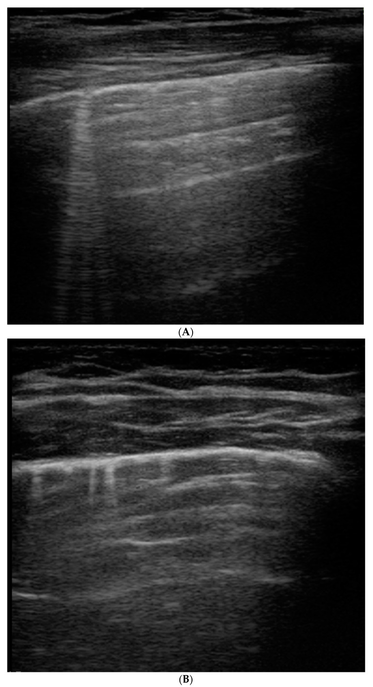
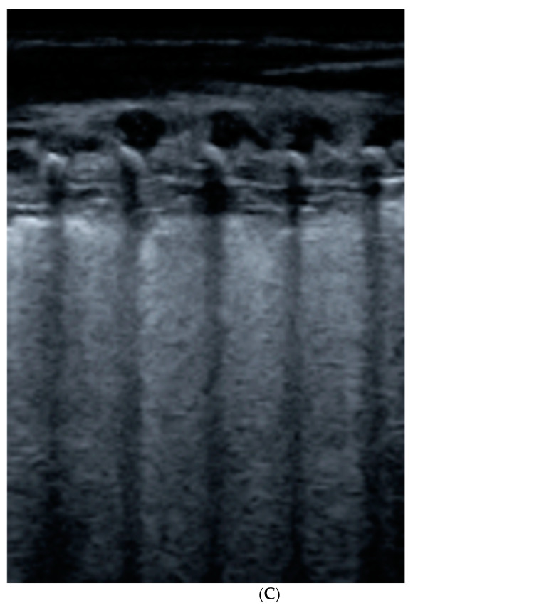
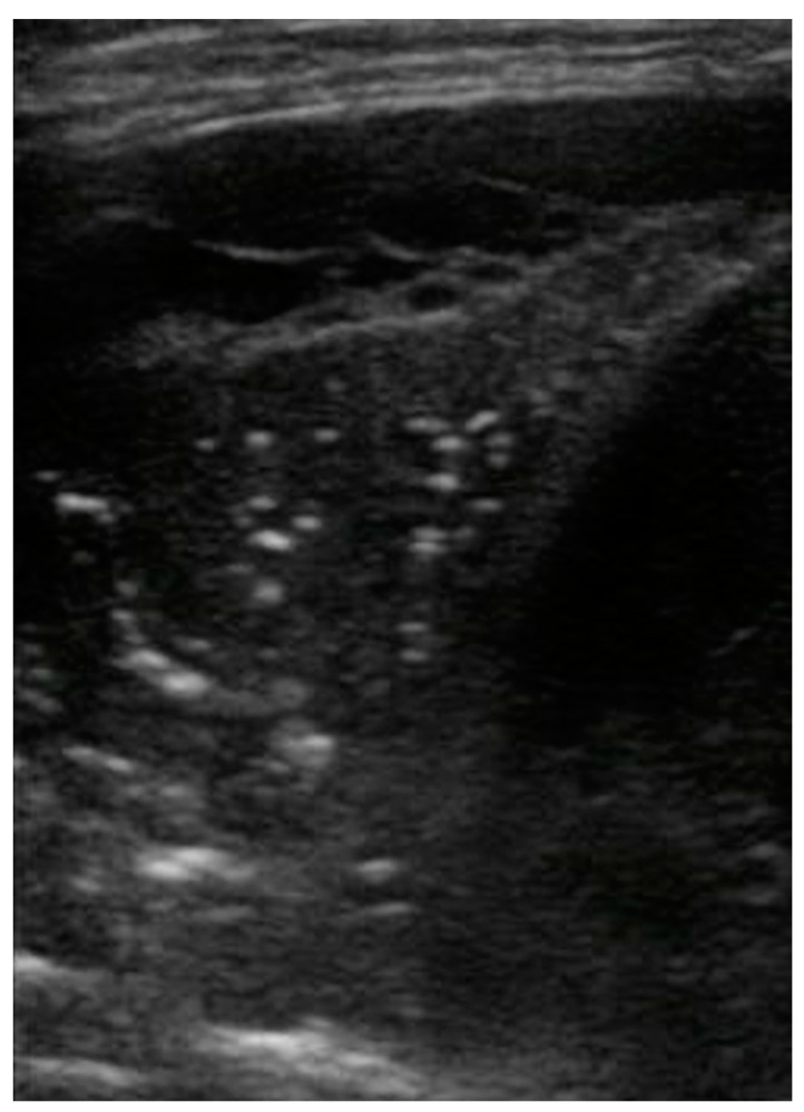
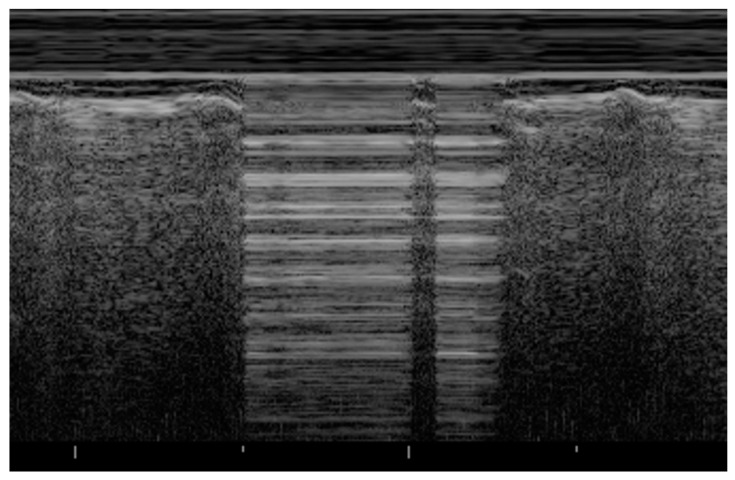
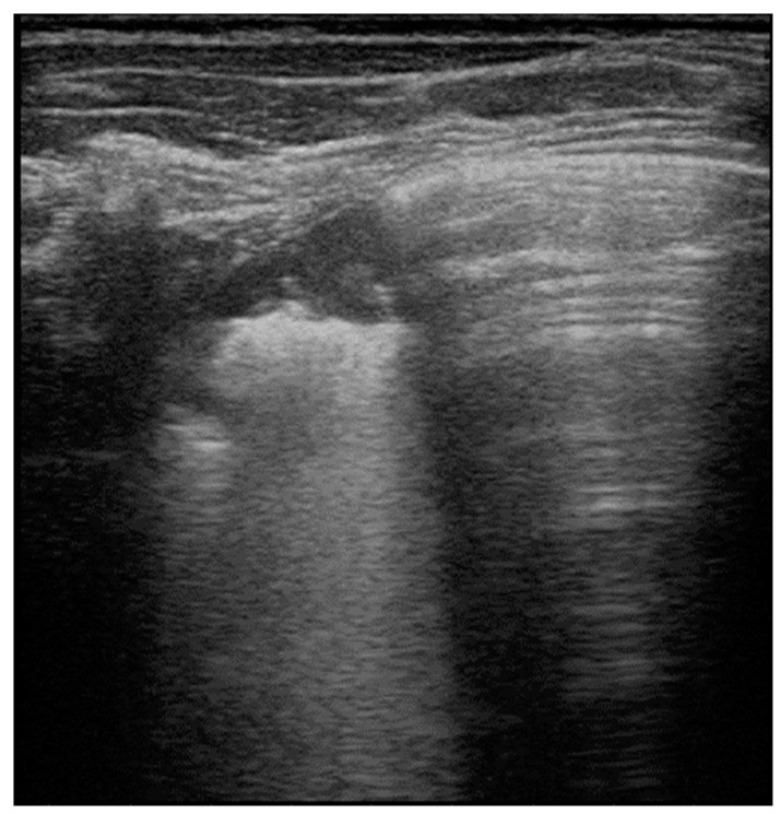
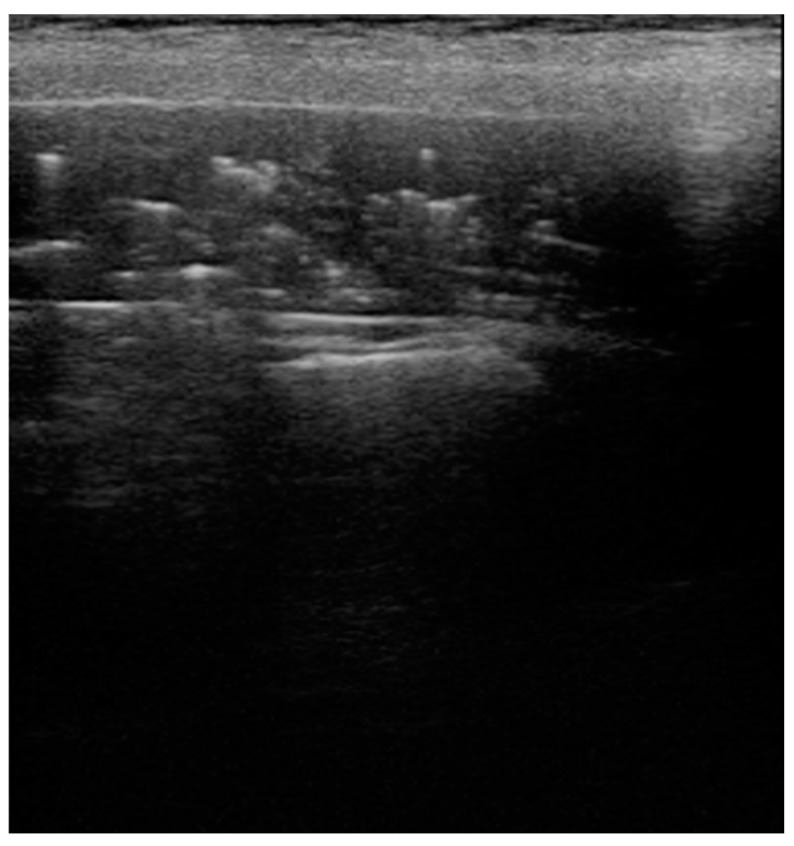
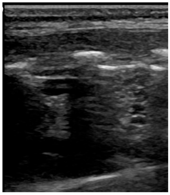
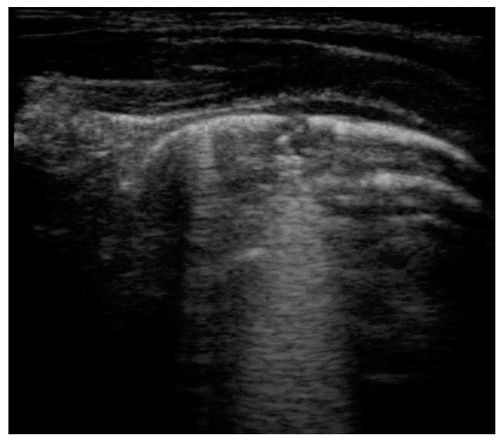
The potential role of ultrasound for the diagnosis of pulmonary diseases is a recent field of research, because, traditionally, lungs have been considered unsuitable for ultrasonography for the high presence of air and thoracic cage that prevent a clear evaluation of the organ. The peculiar anatomy of the pediatric chest favors the use of lung ultrasound (LUS) for the diagnosis of respiratory conditions through the interpretation of artefacts generated at the pleural surface, correlating them to disease-specific patterns. Recent studies demonstrate that LUS can be a valid alternative to chest X-rays for the diagnosis of pulmonary diseases, especially in children to avoid excessive exposure to ionizing radiations. This review focuses on the description of normal and abnormal findings during LUS of the most common pediatric pathologies. Current literature demonstrates usefulness of LUS that may become a fundamental tool for the whole spectrum of lung pathologies to guide both diagnostic and therapeutic decisions.

Keywords: imaging; lung imaging; lung pathology; lung ultrasound; pediatrics; ultrasonography.


Conflict of interest statement

The authors declare no conflict of interest.

Figures

Figure 1
Figure 1
B-lines: vertical hyperechoic artifacts, extending from the pleura that erase normal A-lines (horizontal hyperechoic artifacts). They are the sign of increased interstitial fluid content and thickening of interlobular septae. (A) Deep B-lines, (B) short B-lines, (C) compact B-lines: the number of these artifacts is proportional to the decrease in air content with a tendency to confluence in severe alveolar–interstitial syndrome.
Figure 1
Figure 1
B-lines: vertical hyperechoic artifacts, extending from the pleura that erase normal A-lines (horizontal hyperechoic artifacts). They are the sign of increased interstitial fluid content and thickening of interlobular septae. (A) Deep B-lines, (B) short B-lines, (C) compact B-lines: the number of these artifacts is proportional to the decrease in air content with a tendency to confluence in severe alveolar–interstitial syndrome.
Figure 2
Figure 2
Complex pleural effusion associated with bacterial pneumonia: presence of fibrinous strands and septations (echogenic appearance) in the typical anechoic image related to fluid.
Figure 3
Figure 3
Pneumothorax: evidence of lung point, i.e., the transition point from the typical LUS pattern of PNX to the normal one.
Figure 4
Figure 4
Mycoplasma pneumonia: area of consolidation (lung hepatization) with blurred margins and disappearance of pleural line. Adjacent to the affected area, evidence of normal A-lines, i.e., hyperechoic horizontal lines deeper than visible pleural line, parallel and equidistant from one another that are able to exclude the presence of lung pathologies in the scanned area.
Figure 5
Figure 5
Atelectasis in a patient with neuromuscular disease: presence of static horizontal bronchograms that do not move with respiration.
Figure 6
Figure 6
Congenital pulmonary airway malformations: multiple cystic lesions, not found in other pulmonary pathologies.
Figure 7
Figure 7
Bronchiolitis: small subpleural consolidation in a newborn, expression of a disease moderate in severity.

References

    1. Coley B.D. Chest Sonography in Children: Current Indications, Techniques, and Imaging Findings. Radiol. Clin. N. Am. 2011;49:825–846. doi: 10.1016/j.rcl.2011.06.008. - DOI - PubMed
    1. Kurepa D., Zaghloul N., Watkins L., Liu J. Neonatal Lung Ultrasound Exam Guidelines. J. Perinatol. 2018;38:11–22. doi: 10.1038/jp.2017.140. - DOI - PubMed
    1. Liang H.-Y., Liang X.-W., Chen Z.-Y., Tan X.-H., Yang H.-H., Liao J.-Y., Cai K., Yu J.-S. Ultrasound in Neonatal Lung Disease. Quant. Imaging Med. Surg. 2018;8:535–546. doi: 10.21037/qims.2018.06.01. - DOI - PMC - PubMed
    1. Cao H.-Y., Sorantin E. The basic principles of lung ultrasound. In: Liu J., Sorantin E., Cao H.-Y., editors. Neonatal Lung Ultrasonography. Springer; Dordrecht, The Netherlands: 2018. pp. 1–8.
    1. Iovine E., Nenna R., Bloise S., La Regina D.P., Pepino D., Petrarca L., Frassanito A., Lubrano R., Midulla F. Lung Ultrasound: Its Findings and New Applications in Neonatology and Pediatric Diseases. Diagnostics. 2021;11:625. doi: 10.3390/diagnostics11040652. - DOI - PMC - PubMed

LinkOut - more resources