. 2021 Aug 19;9(8):1066.
doi: 10.3390/healthcare9081066.

Morphological Study of the Effect of Aerobic Exercise on Organs and Arteries in Spontaneously Hypertensive Rats

Affiliations

Morphological Study of the Effect of Aerobic Exercise on Organs and Arteries in Spontaneously Hypertensive Rats

Yingzhe Xiong et al. Healthcare (Basel). .

Abstract

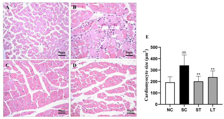
Hypertension is usually accompanied by the impairment of organs and arteries, and seriously threatens human health. Aerobic exercise can effectively prevent and treat hypertension. However, the mechanism of exercise therapy in hypertension is still unclear. In this study, we explored how aerobic exercise effectively reversed the impairment of the heart, kidney, and arteries caused by hypertension through a pathomorphological perspective. Spontaneously hypertensive rats were subjected to fifteen weeks of 45 min and 90 min swimming training without weight, and we then tested the effect of exercise on the morphology and structure of the heart, kidney, iliac artery, and branch of the mesenteric artery. We found that the myocardial fibers became thinner, the cross-sectional area of myocardial cells decreased, and cardiomyocyte edema disappeared after 45 min of aerobic exercise. Additionally, the pathological microstructure of glomeruli and renal tubules were improved. At the same time, aerobic exercise could also reverse the morphology and structure of arteries and mesenteric artery branches in spontaneously hypertensive rats.

Keywords: aerobic exercise; arteries; heart; hypertension; kidney; morphology.


Conflict of interest statement

The authors declare no conflict of interest.

Figures

Figure 1
Figure 1
Taking Samples. (A) Measuring systolic blood pressure of the caudal artery in rats; (B) physical images of the aortic arch; (C) physical images of the iliac artery; (D) physical images of the branch of mesenteric artery.
Figure 2
Figure 2
Cardiomyocyte. NC group (A), SC group (B), ST group (C), LT group (D), and size of cardiomyocytes (E). Data were expressed as the mean ± SD (n = 8 rats/group). ΔΔ p < 0.01 compared with NC; ** p < 0.01 compared with SC.
Figure 3
Figure 3
The aortic arch. NC group (A), SC group (B), ST group (C), and LT group (D).
Figure 4
Figure 4
The kidney. NC group (A), SC group (B), ST group (C), and LT group (D).
Figure 5
Figure 5
The iliac artery. NC group (A), SC group (B), ST group (C), and LT group (D).
Figure 6
Figure 6
The branch of mesenteric artery. NC group (A), SC group (B), ST group (C), and LT group (D).

References

    1. Rizzoni D., Agabiti-Rosei C., Agabiti-Rosei E. Hemodynamic Consequences of Changes in Microvascular Structure. Am. J. Hypertens. 2017;30:939–946. doi: 10.1093/ajh/hpx032. - DOI - PubMed
    1. Yildiz M., Oktay A.A., Stewart M.H., Milani R.V., Ventura H.O., Lavie C.J. Left ventricular hypertrophy and hypertension. Prog. Cardiovasc. Dis. 2020;63:10–21. doi: 10.1016/j.pcad.2019.11.009. - DOI - PubMed
    1. Stanton T., Dunn F.G. Hypertension, Left Ventricular Hypertrophy, and Myocardial Ischemia. Med. Clin. N. Am. 2017;101:29–41. doi: 10.1016/j.mcna.2016.08.003. - DOI - PubMed
    1. Bourdillon M.T., Vasan R.S. A Contemporary Approach to Hypertensive Cardiomyopathy: Reversing Left Ventricular Hypertrophy. Curr. Hypertens. Rep. 2020;22:85. doi: 10.1007/s11906-020-01092-8. - DOI - PubMed
    1. Ellison D., Farrar F.C. Kidney Influence on Fluid and Electrolyte Balance. Nurs. Clin. N. Am. 2018;53:469–480. doi: 10.1016/j.cnur.2018.05.004. - DOI - PubMed

LinkOut - more resources