. 2021 Jul 23;13(8):2517.
doi: 10.3390/nu13082517.

Long-Term Caffeine Intake Exerts Protective Effects on Intestinal Aging by Regulating Vitellogenesis and Mitochondrial Function in an Aged Caenorhabditis Elegans Model

Affiliations

Long-Term Caffeine Intake Exerts Protective Effects on Intestinal Aging by Regulating Vitellogenesis and Mitochondrial Function in an Aged Caenorhabditis Elegans Model

Hyemin Min et al. Nutrients. .

Abstract

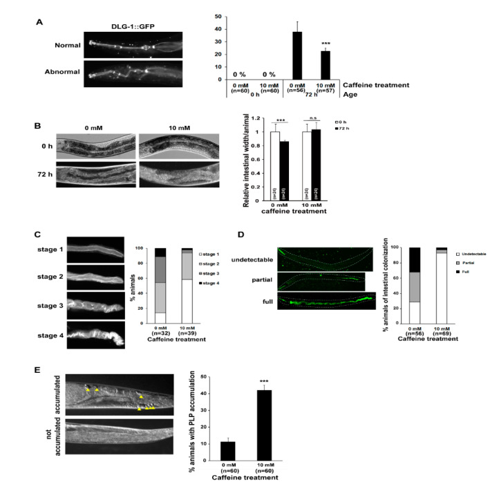
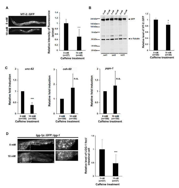
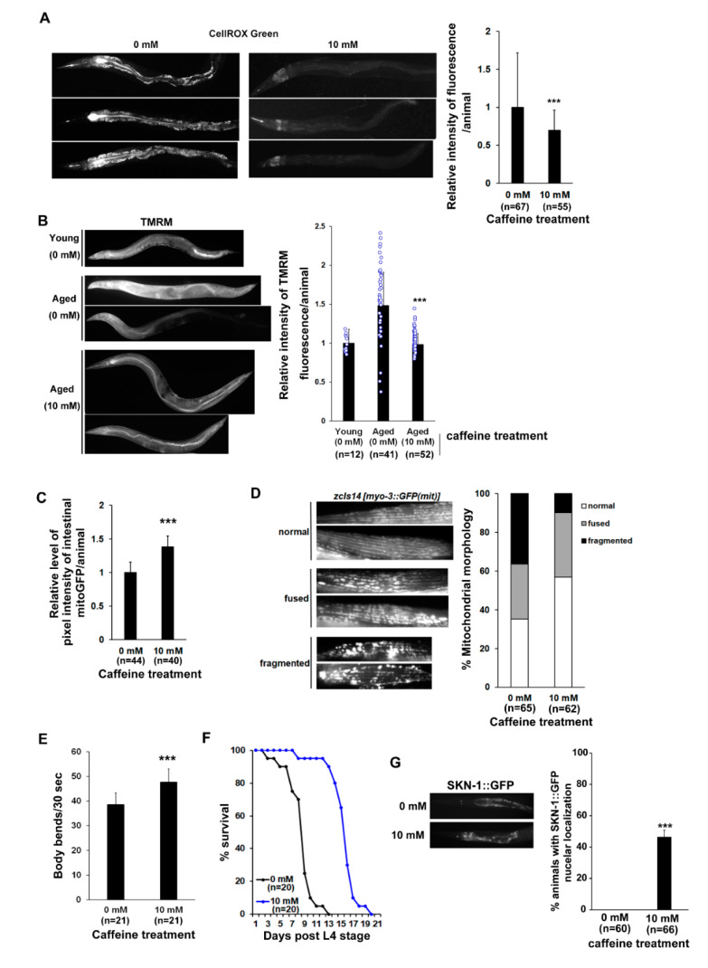
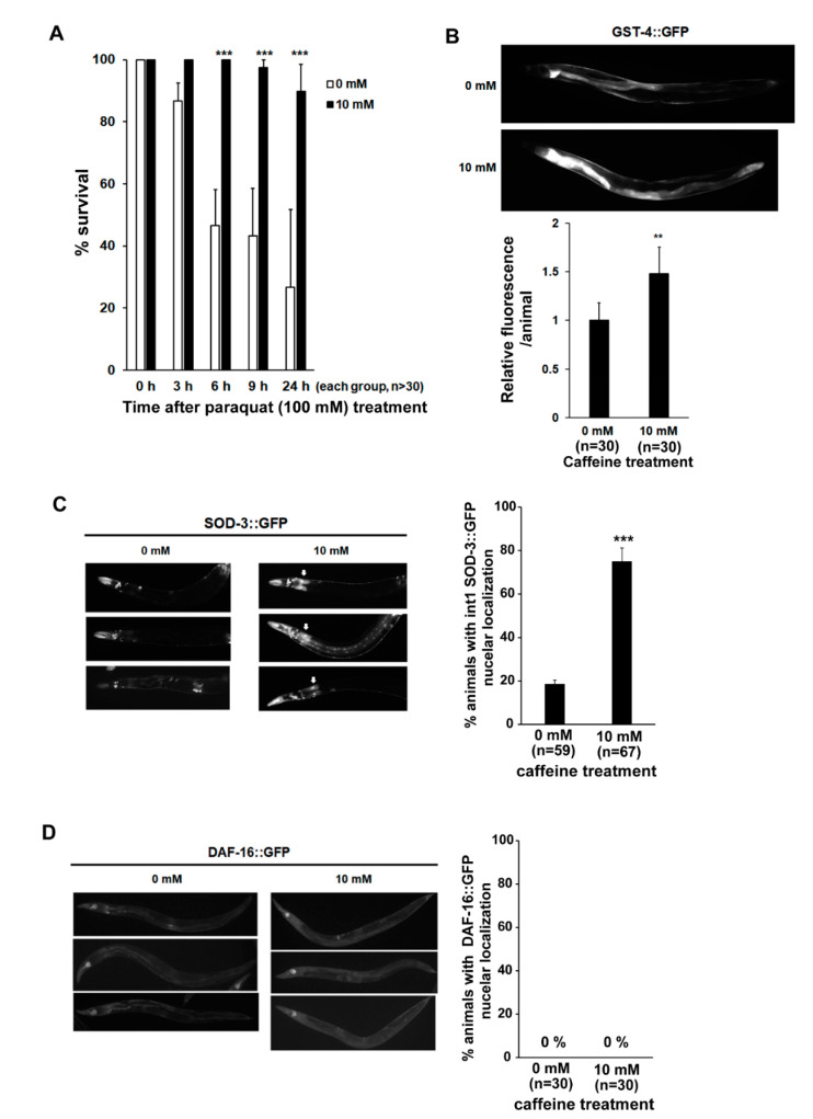
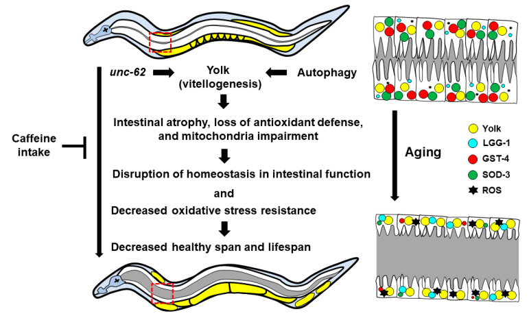
Caffeine, a methylxanthine derived from plants, is the most widely consumed ingredient in daily life. Therefore, it is necessary to investigate the effects of caffeine intake on essential biological activities. In this study, we attempted to determine the possible anti-aging effects of long-term caffeine intake in the intestine of an aged Caenorhabditis elegans model. We examined changes in intestinal integrity, production of vitellogenin (VIT), and mitochondrial function after caffeine intake. To evaluate intestinal aging, actin-5 (ACT-5) mislocalization, lumenal expansion, and intestinal colonization were examined after caffeine intake, and the levels of vitellogenesis as well as the mitochondrial activity were measured. We found that the long-term caffeine intake (10 mM) in the L4-stage worms at 25 °C for 3 days suppressed ACT-5 mislocalization. Furthermore, the level of autophagy, which is normally increased in aging animals, was significantly reduced in these animals, and their mitochondrial functions improved after caffeine intake. In addition, the caffeine-ingesting aging animals showed high resistance to oxidative stress and increased the expression of antioxidant proteins. Taken together, these findings reveal that caffeine may be a potential anti-aging agent that can suppress intestinal atrophy during the progression of intestinal aging.

Keywords: Caenorhabditis elegans; anti-aging; caffeine; intestinal aging; mitochondrial function; oxidative stress response; vitellogenesis.


Conflict of interest statement

The authors declare no conflict of interest.

Figures

Figure 1
Figure 1
Long-term caffeine intake delays intestinal aging during advanced ages of Caenorhabditis elegans: (A) Animals expressing discs large MAGUK scaffold protein 1 (DLG-1):: green fluorescent protein (GFP) were treated with 0 or 10 mM caffeine at the L4 stage at 25 °C for 72 h. The location of DLG-1::GFP in the pharynx was observed at 72 h post-L4 stage at 25 °C. Error bars represent standard deviation (SD). *** p < 0.001 (one-way analysis of variance (ANOVA) with Tukey’s post hoc test); (B) The synchronized wild-type L4-stage animals were treated with 0 or 10 mM caffeine at 25 °C for 72 h. The intestinal atrophy was measured at 72 h post-L4 stage at 25 °C. Error bars represent SD. *** p < 0.001. n.s., not significant (two-way ANOVA with Tukey’s post hoc test); (C) Animals expressing actin 5 (ACT-5)::GFP were treated with 0 or 10 mM caffeine at the L4 stage at 25 °C for 72 h. The type of mislocalization was classified into four stages. The percent distributions of the respective stages in animals fed with 0 or 10 mM caffeine are presented; (D) The synchronized wild-type L4-stage animals were fed with E. coli OP50::GFP, a fluorescent bacteria on 0 or 10 mM caffeine nematode growth medium (NGM) plates at 25 °C for 72 h. The type of bacterial colonization was classified into three categories: (1) undetectable, (2) partial, and (3) full. The percent distributions of the respective categories in animals treated with 0 or 10 mM caffeine are presented; (E) The synchronized wild-type L4-stage animals were treated with 0 or 10 mM caffeine at 25 °C for 72 h. Accumulation of the pseudocoelomic lipoprotein pool (PLP) (indicated by yellow arrowheads) was observed at 72 h in post-L4-stage animals at 25 °C. The percentage of animals with PLP accumulation among the total number of animals is shown. Error bars represent SD. *** p < 0.001 (t-test).
Figure 2
Figure 2
Long-term caffeine intake decreases vitellogenesis in advanced ages of C. elegans: (A) Animals expressing vitellogenin (VIT)-2::GFP were treated with 0 or 10 mM caffeine at the L4 stage at 25 °C for 72 h. The expression of VIT-2::GFP was observed at 72 h in post-L4-stage animals at 25 °C. Error bars represent SD. *** p < 0.001 (t-test); (B) Western blot analysis of VIT-2::GFP protein levels using an anti-GFP antibody in each test condition. α-tubulin was used as the loading control. The relative expression levels of GFP in each condition are shown. GFP band intensity was normalized to that of α-tubulin on the same lane, and the relative levels of GFP were converted to a relative value against that of the animals fed with 0 mM caffeine as 1. Error bars represent SD. * p < 0.05 (t-test); (C) The synchronized wild-type L4-stage animals were treated with 0 or 10 mM caffeine at 25 °C for 72 h. The mRNA levels of unc-62, ceh-60, and pqm-1 in the caffeine-free or caffeine-ingested animals were determined by three independent quantitative reverse transcription-polymerase chain reaction (qRT-PCR) tests using the mRNA level of act-1 in each sample as an internal control for normalization. Error bars represent SD. *** p < 0.001. n.s., not significant (t-test); (D) Animals expressing lgg-1p::GFP::lgg-1 were treated with 0 or 10 mM caffeine at the L4 stage at 25 °C for 72 h; then, we imaged GFP::LGG-1 foci in the intestinal cells at 72 h post-L4-stage animals at 25 °C. The white dotted box represents the GFP::LGG-1 foci in the intestinal cells, and the right panel shows an enlarged image. The graph shows the relative levels of GFP::LGG-1 foci in the intestinal cells after caffeine treatment. Error bars represent SD. *** p < 0.001. n.s., not significant (t-test).
Figure 3
Figure 3
Long-term caffeine intake improves mitochondrial function and motility during advanced ages of C. elegans: (A) Comparison of the mitochondrial reactive oxygen species (ROS) levels between the caffeine-free and caffeine-ingested animals by CellROX Green staining. Wild-type animals were treated with 0 or 10 mM caffeine at the L4 stage at 25 °C for 72 h. The graph shows the relative levels of mitochondrial ROS analyzed by ImageJ. Error bars represent SD. *** p < 0.001 (t-test); (B) Comparison of the mitochondrial membrane potential (MMP) in young (0 mM) and aged (0 mM; 10 mM) animals using tetramethylrhodamine methyl ester (TMRM) staining. In the aged groups, wild-type animals were treated with 0 or 10 mM caffeine at the L4 stage at 25 °C for 72 h. In the young (0 mM) group, wild-type animals were treated with 0 mM caffeine at the L4 stage at 25 °C for 24 h. The TMRM fluorescence was quantified for each test condition by ImageJ. The graph shows the relative levels of MMP. Error bars represent SD. *** p < 0.001 (one-way ANOVA with Tukey’s post hoc test); (C) Comparison of intestinal mitochondrial activity in ges-1p::GFP(mit) transgenic animal expressing ges-1 promoter driven GFP. The transgenic animals were treated with 0 or 10 mM caffeine at the L4 stage at 25 °C for 72 h. The graph shows the relative levels of fluorescence intensity as analyzed by ImageJ. Error bars represent SD. *** p < 0.001 (t-test); (D) The mitochondrial morphology was analyzed using the SJ4103 transgenic animal expressing a mitochondrial-targeted GFP under the control of the muscle-specific myo-3 promoter. The transgenic animals were treated with 0 or 10 mM caffeine at the L4 stage at 25 °C for 72 h. The graph indicates the percentage of animals with muscle mitochondria classified into three categories: (1) normal, (2) fused, and (3) fragmented; (E) Comparison of body bending in caffeine-free diet animals and caffeine-ingested animals at advanced ages. Wild-type animals were treated with 0 or 10 mM caffeine at the L4 stage at 25 °C for 72 h. Error bars represent SD. *** p < 0.001 (t-test); (F) Comparison of the survival rates between the caffeine-free diet animals and caffeine-ingested animals. Wild-type animals were treated with 0 or 10 mM caffeine at the L4 stage until dead at 25 °C; (G) Animals expressing skinhead 1 (SKN-1)::GFP were treated with 0 or 10 mM caffeine at the L4 stage at 25 °C for 72 h. The expression levels of SKN-1::GFP were observed at 72 h post-L4-stage animals at 25 °C. Error bars represent SD. *** p < 0.001 (t-test).
Figure 4
Figure 4
Long-term caffeine intake exerts a protective effect on oxidative stress in advanced ages of C. elegans: (A) Percentage survival rates of caffeine-free diet and caffeine-ingested animals were analyzed under paraquat (100 mM)-induced oxidative stress condition. Error bars represent SD. *** p < 0.001 (two-way ANOVA with Tukey’s post hoc test); (B) Animals expressing glutathione S-transferase 4 (GST-4)::GFP were treated with 0 or 10 mM caffeine at the L4 stage at 25 °C for 72 h. The expression of GST-4::GFP was observed at 72 h post-L4-stage animals at 25 °C. The graph shows the relative fluorescence intensity analyzed by ImageJ. Error bars represent SD. ** p < 0.01 (t-test); (C) Animals expressing superoxide dismutase 3 (SOD-3)::GFP were treated with 0 or 10 mM caffeine at the L4 stage at 25 °C for 72 h. The expression levels of SOD-3::GFP were observed at 72 h post-L4-stage animals at 25 °C. The graph indicates the percentage of animals with SOD-3::GFP nuclear localization in the intestinal cells. Error bars represent SD. *** p < 0.001 (t-test); (D) Animals expressing DAF-16::GFP were treated with 0 or 10 mM caffeine at the L4 stage at 25 °C for 72 h. The expression levels of DAF-16::GFP were observed at 72 h post-L4-stage animals at 25 °C. The graph indicates the percentage of animals with DAF-16::GFP nuclear localization in the intestinal cells.
Figure 5
Figure 5
A working model to explain the protective effects of long-term caffeine intake on intestinal aging C. elegans at advanced ages. Long-term caffeine intake reduces vitellogenesis via regulating the expression of unc-62 and autophagy. The decrease in vitellogenesis in response to caffeine intake delays the intestinal atrophy along with improving the mitochondrial function and antioxidant defense. Maintaining homeostasis of intestinal function via caffeine intake supports the health and lifespan in C. elegans at advanced ages.

References

    1. Min H., Kawasaki I., Gong J., Shim Y.H. Caffeine induces high expression of cyp-35A family genes and inhibits the early larval development in Caenorhabditis elegans. Mol. Cells. 2015;38:236–242. doi: 10.14348/molcells.2015.2282. - DOI - PMC - PubMed
    1. Du X., Guan Y., Huang Q., Lv M., He X., Yan Y., Hayashi S., Fang C., Wang X., Sheng J. Low concentrations of caffeine and its analogs extend the lifespan of Caenorhabditis elegans by modulating IGF-1-Like pathway. Front. Aging Neurosci. 2018;10:211. doi: 10.3389/fnagi.2018.00211. - DOI - PMC - PubMed
    1. Li H., Roxo M., Cheng X., Zhang S., Cheng H., Wink M. Pro-oxidant and lifespan extension effects of caffeine and related methylxanthines in Caenorhabditis elegans. Food Chem. X. 2019;1:100005. doi: 10.1016/j.fochx.2019.100005. - DOI - PMC - PubMed
    1. Min H., Youn E., Shim Y.H. Maternal caffeine intake disrupts eggshell integrity and retards larval development by reducing yolk production in a Caenorhabditis elegans model. Nutrients. 2020;12:1334. doi: 10.3390/nu12051334. - DOI - PMC - PubMed
    1. Sutphin G.L., Bishop E., Yanos M.E., Moller R.M., Kaeberlein M. Caffeine extends life span, improves healthspan, and delays age-associated pathology in Caenorhabditis elegans. Logev. Healthspan. 2012;1:9. doi: 10.1186/2046-2395-1-9. - DOI - PMC - PubMed

LinkOut - more resources