Review
. 2021 Aug 4;22(16):8370.
doi: 10.3390/ijms22168370.

Reviewing the Significance of Blood-Brain Barrier Disruption in Multiple Sclerosis Pathology and Treatment

Affiliations
Review

Reviewing the Significance of Blood-Brain Barrier Disruption in Multiple Sclerosis Pathology and Treatment

Rodica Balasa et al. Int J Mol Sci. .

Abstract

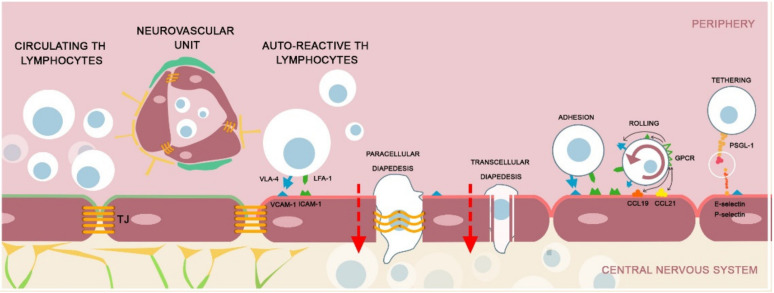
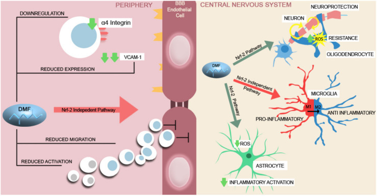
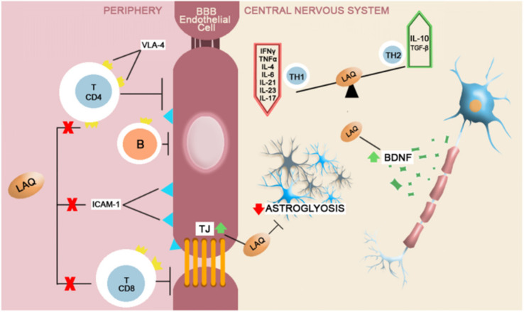
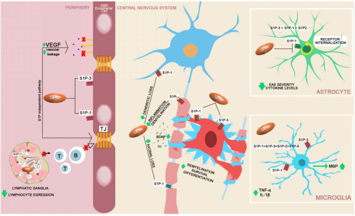
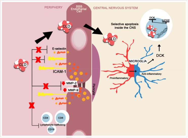
The disruption of blood-brain barrier (BBB) for multiple sclerosis (MS) pathogenesis has a double effect: early on during the onset of the immune attack and later for the CNS self-sustained 'inside-out' demyelination and neurodegeneration processes. This review presents the characteristics of BBB malfunction in MS but mostly highlights current developments regarding the impairment of the neurovascular unit (NVU) and the metabolic and mitochondrial dysfunctions of the BBB's endothelial cells. The hypoxic hypothesis is largely studied and agreed upon recently in the pathologic processes in MS. Hypoxia in MS might be produced per se by the NVU malfunction or secondary to mitochondria dysfunction. We present three different but related terms that denominate the ongoing neurodegenerative process in progressive forms of MS that are indirectly related to BBB disruption: progression independent of relapses, no evidence of disease activity and smoldering demyelination or silent progression. Dimethyl fumarate (DMF), modulators of S1P receptor, cladribine and laquinimode are DMTs that are able to cross the BBB and exhibit beneficial direct effects in the CNS with very different mechanisms of action, providing hope that a combined therapy might be effective in treating MS. Detailed mechanisms of action of these DMTs are described and also illustrated in dedicated images. With increasing knowledge about the involvement of BBB in MS pathology, BBB might become a therapeutic target in MS not only to make it impenetrable against activated immune cells but also to allow molecules that have a neuroprotective effect in reaching the cell target inside the CNS.

Keywords: blood-brain barrier; disease modifying therapies progression; impermeability; multiple sclerosis.


Conflict of interest statement

The authors declare no conflict of interest.

Figures

Figure 1
Figure 1
A brief schematic presentation of the pathophysiology of MS regarding the disruption of the BBB. The NVU is composed of endothelial cells, astrocyte end-feet and pericytes [9]. The endothelial cells are interconnected by TJs [10,11]. In healthy individuals, the integrity of the BBB maintains the immune circulating cells in the peripheral blood compartment. In MS, the auto-reactive Th cells will extravasate across the vascular endothelium in a complex manner. Tethering: The peripheral lymphocytes express P-selectin glycoprotein ligand-1 (PSGL-1) that interact with the ligand molecules expressed on the endothelial cells (E and P-selectins). Rolling: The contact between the lymphocyte and the endothelium is preceded by the rolling beside the vessel wall–a transient, reversible process. The endothelial cells express various chemokines (CCL21, CCL19) that will activate the G protein coupled receptor (GPCR) on the surface of the lymphocyte and stimulate the expression of integrins, very late antigen-4 (VLA-4) and lymphocyte function associated antigen 1 (LFA-1) [21,22,23,24]. Adhesion: The lymphocyte will adhere to the endothelial cells by coupling the surface adhesion molecules (VLA-4 and LFA-1) with the endothelial cell receptors (VCAM-1 and ICAM-1). After coupling, the lymphocytes will transverse the BBB by transcellular or paracellular pathways [25]. The activated T lymphocytes have the capacity to alter the inflamed BBB and create a transendothelial pore by modelling the caveolin-1 and transverse by a transcellular pathway. In the paracellular breakthrough, the T lymphocytes remodel the TJs by altering the connective molecules (occludin, claudin) and create a permissive ‘fenestration’ [26,27,28,29].
Figure 2
Figure 2
A brief schematic representation of DMF’s mechanism of action. In the peripheral compartment, DMF induces changes in the immune response by decreasing the activation and migration of Th lymphocytes. It further alters the transendothelial migration across the BBB, reduces the expression of the adhesion molecules, such as VCAM-1, and downregulates the α4 integrin on the lymphocyte surface [81,82,84,85,88,89]. Inside the CNS, DMF carries Nrf-2 dependent neuroprotective effects upon the neurons and oligodendrocytes by increasing the ROS resistance of the neurons and glial cells and upon the astrocytes by decreasing the proinflammatory cytokine secretion and intracellular ROS production [86,90,91]. In proinflammatory activated microglia, DMF reduces the production of proinflammatory mediators and stimulates the shift from a M1 proinflammatory state to a M2, anti-inflammatory state [86,92].
Figure 3
Figure 3
A brief schematic representation of Laquinimod’s mechanism of action. In the peripheral compartment, Laquinimod inhibits the lymphocytic differentiation for CD4+ and CD8+ Th cells, limits the B cells passage and the lymphocytic endothelial adhesion by down-regulating VLA-4 and ICAM-1 [97,101,102,103]. The neuroprotective effects of laquinimod are dependent on BDNF up-modulation with secondary reduced axonal loss. It also reduces astroglyosis and oligodendrocyte apoptosis and reduces the expression of proinflammatory Th1 cytokines, while augmenting the T2 anti-inflammatory response [104,105,106].
Figure 4
Figure 4
A brief schematic representation of fingolimod’s mechanism of action. In the periphery, fingolimod (sphingosine phosphate receptor modulator–SPM) acts upon the sphingosine 1-phosphate receptors (S1P1, S1P2, S1P3, S1P4, S1P5) [101]. In the lymphatic ganglia, SPM antagonize the S1P1 expression and blocks the lymphocytic egression into the circulation (T and B). SPM blocks the expression of S1P1 and S1P3 on the surface of the endothelial cells, reducing lymphocyte transmigration. Immune cell passage at the level of the BBB is reduced secondary VEGF reduced expression [109,110,111,112]. Inside the CNS the S1P receptors are expressed by the majority of neural cell lineages [113]. S1P1 and S1P5 modulation enhances oligodendrocyte function, survival, differentiation and secondarily boosts the remyelination [114,115]. Neuronal function is directly sustained by modulation of S1P1 and S1P3 receptors and by the up-regulation of BNDF expression, with neuroprotective effects [113,116]. The astrocytes, secondary to S1P1 modulation were proven to decrease EAE severity in murine studies and decrease the levels of proinflammatory cytokines [117,118]. fingolimod and SPM reduce proinflammatory cytokine production from microglia and increase the secretion of myelin basic protein (MBP) after a demyelinating event, promoting remyelination [119,120].
Figure 5
Figure 5
A brief schematic representation of CLD’s mechanism of action. In the peripheral compartment, CLD reduces the expression of adhesion molecules, ICAM-1 and E-selectin and reduces the expression of MMP-2 and -9 [136,137,138,139], thus inhibiting the lymphocyte transition into the CNS. CLD is internalized into the cells and undergoes specific phosphorylation by deoxycytidine kinase (DCK). Immune cells, the lymphocytes being the most susceptible, contain reduced levels of phosphatases 5-nucleotidases (5-NTASE), which will only partially dephosphorylate the accumulated CLD, leading to selected apoptosis and not cellular death (ratio of DCK to 5-NTASE) [140,141]. In MS, CLD selectively reduces Th and B lymphocyte numbers and trafficking from the periphery [142]. CLD reduces the activation of proinflammatory microglia (M1) and shifts the activity towards an anti-inflammatory (M2) phenotype [138].

References

    1. Kobelt G., Thompson A., Berg J., Gannedahl M., Eriksson J., MSCOI Study Group. European Multiple Sclerosis Platform New insights into the burden and costs of multiple sclerosis in Europe. Mult. Scler. 2017;23:1123–1136. doi: 10.1177/1352458517694432. - DOI - PMC - PubMed
    1. Spencer J.I., Bell J.S., DeLuca G.C. Vascular pathology in multiple sclerosis: Reframing pathogenesis around the blood-brain barrier. J. Neurol. Neurosurg. Psychiatry. 2018;89:42–52. doi: 10.1136/jnnp-2017-316011. - DOI - PubMed
    1. Titus H.E., Chen Y., Podojil J.R., Robinson A.P., Balabanov R., Popko B., Miller S.D. Pre-clinical and Clinical Implications of “Inside-Out” vs. “Outside-In” Paradigms in Multiple Sclerosis Etiopathogenesis. Front. Cell Neurosci. 2020;14:599717. doi: 10.3389/fncel.2020.599717. - DOI - PMC - PubMed
    1. Goodin D.S., Reder A.T., Bermel R.A., Cutter G.R., Fox R.J., John G.R., Lublin F.D., Luccinetti C.F., Miller A.E., Pelletier D., et al. Relapses in multiple sclerosis: Relationship to disability. Mult. Scler. Relat. Disord. 2016;6:10–20. doi: 10.1016/j.msard.2015.09.002. - DOI - PubMed
    1. Leray E., Yaouanq J., Le Page E., Coustans M., Laplaud D., Oger J., Edan G. Evidence for a two-stage disability progression in multiple sclerosis. Brain. 2010;133:1900–1913. doi: 10.1093/brain/awq076. - DOI - PMC - PubMed

MeSH terms