. 2021 Aug 5;22(16):8421.
doi: 10.3390/ijms22168421.

Stimulating GABAergic Neurons in the Nucleus Accumbens Core Alters the Trigeminal Neuropathic Pain Responses in a Rat Model of Infraorbital Nerve Injury

Affiliations

Stimulating GABAergic Neurons in the Nucleus Accumbens Core Alters the Trigeminal Neuropathic Pain Responses in a Rat Model of Infraorbital Nerve Injury

Jaisan Islam et al. Int J Mol Sci. .

Abstract

The nucleus accumbens core (NAcc) is an important component of brain reward circuitry, but studies have revealed its involvement in pain circuitry also. However, its effect on trigeminal neuralgia (TN) and the mechanism underlying it are yet to be fully understood. Therefore, this study aimed to examine the outcomes of optogenetic stimulation of NAcc GABAergic neurons in an animal model of TN. Animals were allocated into TN, sham, and control groups. TN was generated by infraorbital nerve constriction and the optogenetic virus was injected into the NAcc. In vivo extracellular recordings were acquired from the ventral posteromedial nucleus of the thalamus. Alterations of behavioral responses during stimulation "ON" and "OFF" conditions were evaluated. In vivo microdialysis was performed in the NAcc of TN and sham animals. During optogenetic stimulation, electrophysiological recordings revealed a reduction of both tonic and burst firing activity in TN animals, and significantly improved behavioral responses were observed as well. Microdialysis coupled with liquid chromatography/tandem mass spectrometry analysis revealed significant alterations in extracellular concentration levels of GABA, glutamate, acetylcholine, dopamine, and citrulline in NAcc upon optic stimulation. In fine, our results suggested that NAcc stimulation could modulate the transmission of trigeminal pain signals in the TN animal model.

Keywords: VPM thalamus; microdialysis; nucleus accumbens core; optogenetics; trigeminal neuralgia.


Conflict of interest statement

The authors declare no conflict of interest. The funders had no role in the design of the study; in the collection, analyses, or interpretation of data; in the writing of the manuscript, or in the decision to publish the results.

Figures

Figure 1
Figure 1
Changes in pain behaviors following ION ligation. Behavioral scores of the TN, sham, and control groups (AH). (A) Results from the air-puff test for the ipsilateral trigeminal facial area. (B) Cold allodynia score results for the ipsilateral facial area with acetone drops. (C) Mechanical allodynia (von Frey test) results for the ipsilateral facial side. (DH) Results of the open field test: (D) number of explored areas, (E) rearing events, (F) grooming time, (G) active time, and (H) distance traveled. **, p < 0.01; ***, p < 0.001; ****, p < 0.0001, significant difference determined using an analysis of variance. Data are represented as means ± SD.
Figure 2
Figure 2
Extracellular recording data in vivo. (A) Schematic diagram showing extracellular recording area. (B) Evoked firing rates in VPM neurons in TN (n = 32) animals versus sham (n = 32) and control (n = 4) animals (****, p < 0.0001, significant difference determined using unpaired t-tests). (C,D) In vivo recordings of the VPM thalamus from TN-Opto animals. (C) Findings for the TN-Opto-CGRP (n = 8) group. (D) Findings for the TN-Opto-PBS (n = 8) group. A decrease in firing output (spikes/s) was observed during the stimulation “ON” period. (E,F) No changes in the VPM thalamus firing rates were observed in the TN-Null-CGRP (n = 8) or TN-Null-PBS (n = 8) groups (two-way analysis of variance *, p < 0.05; **, p < 0.01; ***, p < 0.001). (G,H) Changes in the burst firing rates of the TN animals following optogenetic stimulation. A significant reduction was observed in the TN-Opto-CGRP (G, blue line) and TN-Opto-PBS (H, blue line) groups. ***, p < 0.001, analysis of variance. (I,J) Peri-event raster histogram of VPM neuron responses show spike traces (above), raster traces (middle), and rate histogram (below) in the TN-Opto-CGRP (I) and TN-Opto-PBS (J) groups in optic stimulation “ON and “OFF” states. Bin size = 50 ms. VPM thalamic responses were decreased during optic stimulation (blue area).
Figure 3
Figure 3
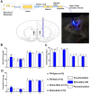
Attenuation of hyperalgesia by optogenetic stimulation. (A) Optogenetic stimulation setup and area. (BD) Changes in behavioral responses following blue laser stimulation: results from the air-puff test (B), cold allodynia score test (C), and von Frey test (D) of TN-Opto (n = 12) (a), TN-Null (n = 12) (b), Sham-Opto (n = 12) (c), and Sham-Null (n = 12) (d) animal groups, respectively. Only the TN-Opto group exhibited significant changes in behavioral scores during the stimulation “ON” condition. No significant changes in behavioral scores were observed in other animal groups during the stimulation “ON” condition. *, p < 0.05; **, p < 0.01, significant differences determined using an analysis of variance. Data are represented as means ± SD.
Figure 4
Figure 4
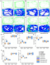
Alteration of behavioral responses in the open field test by optogenetic stimulation. (AD) Trajectory and exploration representations of the TN-Opto, TN-Null, Sham-Opto, and Sham-Null groups, respectively, during stimulation “OFF” and “ON” conditions. (E) Numbers of explored areas, (F) rearing events, (G) grooming time, (H) active time, and (I) distance traveled. In all the behavioral paradigms, the TN-Opto group exhibited significant alterations with the stimulation “ON” condition but no significant changes in behavioral scores were observed in other animal groups with the stimulation “ON” condition. *, p < 0.05; **, p < 0.01, significant differences determined using unpaired t-test.
Figure 5
Figure 5
Effect of optogenetic stimulation on the extracellular concentration level of GABA, glutamate, dopamine, acetylcholine, and citrulline in NAcc. (A) Microdialysis setup and procedure of microdialysate collection. Extracellular conc. level (ng/mL) of GABA (B), glutamate (C), dopamine (D), acetylcholine (E), and citrulline (F) throughout the 60 min duration (pre-stimulation: 0–20 min; stimulation “ON”: 20–40 min; post-stimulation: 40–60 min). In both TN-Opto (n = 4) and Sham-Opto (n = 4) groups, alterations in the conc. levels of all the five neurotransmitters were observed during optogenetic stimulation but not in the TN-Null (n = 4) and Sham-Null (n = 4) animal groups. *, p < 0.05; **, p < 0.01; ***; p < 0.001, significant differences determined using an analysis of variance. Data are represented as means ± SD.
Figure 6
Figure 6
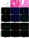
Viral expression, immunostaining of cannula placement, and neuronal activation in the NAcc. (A) H&E staining of brain tissue section showing the anatomical location of NAcc. (B) H&E staining of brain tissue section showing the location of optic fiber and guide cannula placement in NAcc. Scale bar = 50 µm. (CE) Optogenetic viral expression in the NAcc of TN-Opto animals. (FH) Null viral expression in the NAcc of TN-Null animals. (C,F) EYFP, (D,G) DAPI, and (E,H) merged. Scale bar = 200 µm. (IK) Colocalization of staining for c-Fos and GABA by double immunofluorescence labeling in optogenetic virus-injected animal. (LN) Colocalization of staining for c-Fos and GABA by double immunofluorescence labeling in null virus-injected animal. Double immunoreactivity for GABA (I,L), c-Fos (J,M), and merged c-Fos and GABA (K,N) in NAcc. Scale bar = 50 µm.

References

    1. Idänpään-Heikkilä J.J., Guilbaud G. Pharmacological studies on a rat model of trigeminal neuropathic pain: Baclofen, but not carbamazepine, morphine or tricyclic antidepressants, attenuates the allodynia-like behaviour. Pain. 1999;79:281–290. doi: 10.1016/S0304-3959(98)00172-9. - DOI - PubMed
    1. Islam J., Elina K., Oh B.H., Kim S., Hyun S.-H., Park Y.S. Optogenetic stimulation of the motor cortex alleviates neuropathic pain in rats of infraorbital nerve injury with/without CGRP knock-down. J. Headache Pain. 2020;21:1–13. doi: 10.1186/s10194-020-01174-7. - DOI - PMC - PubMed
    1. Henderson J.M., Lad S.P. Motor cortex stimulation and neuropathic facial pain. Neurosurg. Focus. 2006;21:1–4. doi: 10.3171/foc.2006.21.6.9. - DOI - PubMed
    1. Robison A.J., Nestler E.J. Transcriptional and epigenetic mechanisms of addiction. Nat. Rev. Neurosci. 2011;12:623–637. doi: 10.1038/nrn3111. - DOI - PMC - PubMed
    1. Harris H.N., Peng Y.B. Evidence and explanation for the involvement of the nucleus accumbens in pain processing. Neural Regen. Res. 2020;15:597. doi: 10.4103/1673-5374.266909. - DOI - PMC - PubMed

MeSH terms