. 2021 Aug 5;22(16):8434.
doi: 10.3390/ijms22168434.

Somatostatin and Astroglial Involvement in the Human Limbic System in Alzheimer's Disease

Affiliations

Somatostatin and Astroglial Involvement in the Human Limbic System in Alzheimer's Disease

Melania Gonzalez-Rodriguez et al. Int J Mol Sci. .

Abstract

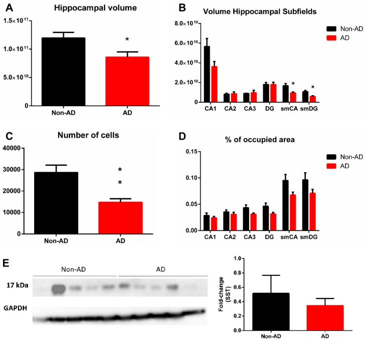
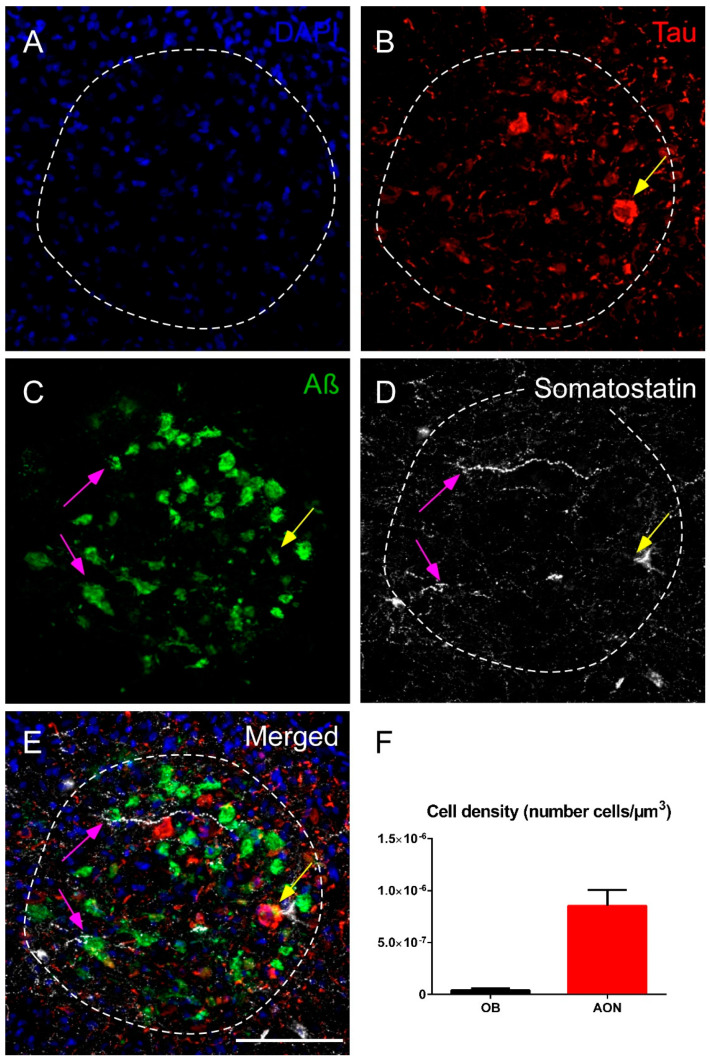
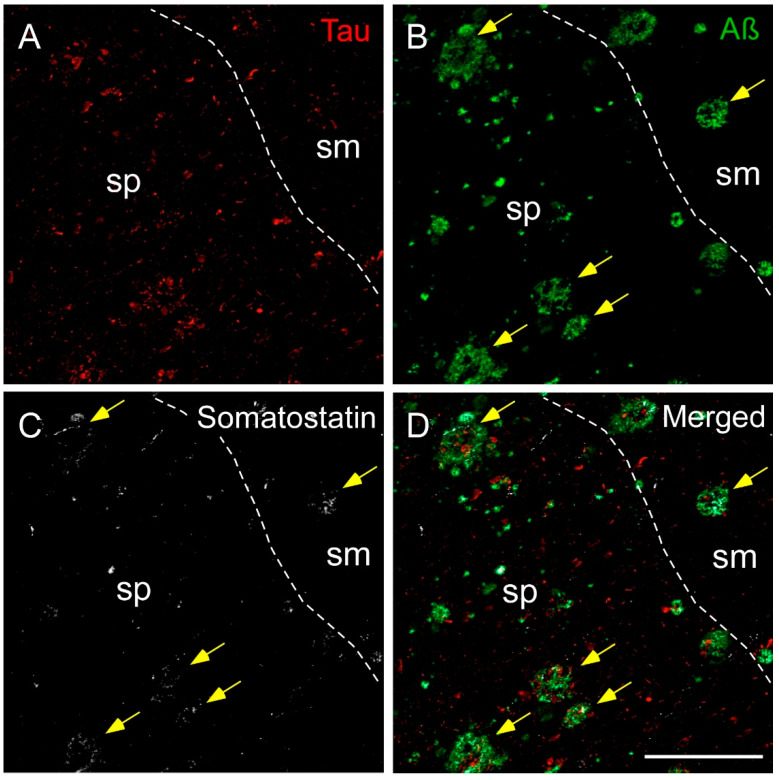
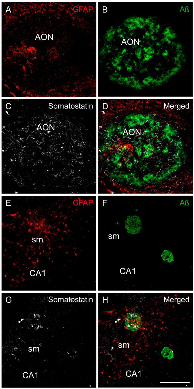
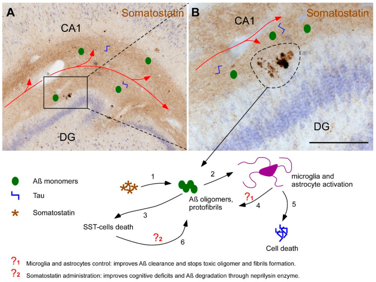
Alzheimer's disease (AD) is the most prevalent neurodegenerative disease in the elderly. Progressive accumulation of insoluble isoforms of amyloid-β peptide (Aβ) and tau protein are the major neuropathologic hallmarks, and the loss of cholinergic pathways underlies cognitive deficits in patients. Recently, glial involvement has gained interest regarding its effect on preservation and impairment of brain integrity. The limbic system, including temporal lobe regions and the olfactory bulb, is particularly affected in the early stages. In the early 1980s, the reduced expression of the somatostatin neuropeptide was described in AD. However, over the last three decades, research on somatostatin in Alzheimer's disease has been scarce in humans. Therefore, the aim of this study was to stereologically quantify the expression of somatostatin in the human hippocampus and olfactory bulb and analyze its spatial distribution with respect to that of Aβ and au neuropathologic proteins and astroglia. The results indicate that somatostatin-expressing cells are reduced by 50% in the hippocampus but are preserved in the olfactory bulb. Interestingly, the coexpression of somatostatin with the Aβ peptide is very common but not with the tau protein. Finally, the coexpression of somatostatin with astrocytes is rare, although their spatial distribution is very similar. Altogether, we can conclude that somatostatin expression is highly reduced in the human hippocampus, but not the olfactory bulb, and may play a role in Alzheimer's disease pathogenesis.

Keywords: Alzheimer’s disease; astroglia; hippocampus; olfactory bulb; somatostatin; stereology.


Conflict of interest statement

The authors declare no conflict of interest.

Figures

Figure 1
Figure 1
Immunohistochemistry staining for somatostatin in the olfactory bulb. Somatostatin is present in all regions within the olfactory bulb (A) with a striking presence in the anterior olfactory nucleus (B), and expression was low in the GrL and within the Mi, IPL, EPL and GL external layers in non-AD cases (C). Similarly, SST-cells did not show neurodegeneration features in AD cases and were mainly placed in the anterior olfactory nucleus (D). In addition, lower intensity of labeling in SST -expressing fibers was observable in the rest of olfactory layers in AD cases (E). Note that the number of somatostatin-expressing cells was very low compared with the number of somatostatin fibers. GrL; granule cell layer, Mi; mitral cell layer, IPL; internal plexiform layer, EPL; external plexiform layer, GL; glomerular layer. Scale bar A, 500 µm; B,C, 200 µm.
Figure 2
Figure 2
Quantification of volume and somatostatin expression in the olfactory bulb. Neither the olfactory bulb nor the anterior olfactory nucleus showed variation in volume (A,B). The results also indicated no difference in either the number of somatostatin-expressing cells (C,D) or the area fraction occupied by somatostatin fibers (E,F). In addition, Western blot quantifications confirmed no change in somatostatin expression in the olfactory bulb (G). For volume, number of cells and somatostatin fibers quantification N = 10 formalin-fixed cases (AD = 5 and non-AD = 5) and 4 sections per case were analyzed. Western blot quantification was performed using N = 20 fresh-frozen cases, AD = 10 and non-AD = 10. See Table 1 and supplementary stereological data for further information.
Figure 3
Figure 3
Immunohistochemistry staining of somatostatin in the hippocampus. Mosaic reconstruction of the non-AD human hippocampus showing somatostatin distribution (A). In non-AD cases, somatostatin-expressing cells were mainly located in either the stratum oriens in CA regions (B) or within the hilus in the DG (C) and somatostatin fibers were mainly localized to the molecular layers of CA-DG regions (D). The major hallmarks of somatostatin in AD brains were dystrophic neurites (E) and aggregates of fibers and cell debris mainly in the molecular layers of CA (F) and DG (G). CA, cornus amonis; DG, dentate gyrus; Sub, subiculum; so, stratum oriens; sp, stratum piramidale; sl, stratum lacunosum; sr, stratum radiatum; sm, stratum moleculare. Scale bar A, 500 µm; BG, 160 µm.
Figure 4
Figure 4
Quantification of volume and somatostatin expression in the hippocampus. Hippocampal volume was reduced in AD (A), and the molecular layers of CA and DG regions were specifically involved (B). The number of somatostatin-expressing cells was diminished in the whole hippocampus (C), but only a tendency (not significant) was observed in the areas occupied by somatostatin fibers (D). Western blot quantification did not show a significant reduction in somatostatin expression in the hippocampus (E). For volume, number of cells and somatostatin fibers quantification N = 10 formalin-fixed cases (AD = 5 and non-AD = 5) and 4 sections per case were analyzed. Western blot quantification was performed using N = 10 fresh-frozen cases, AD = 5 and non-AD = 5. See Table 1 and supplementary stereological data for further information. * Corresponds to p-value < 0.05.
Figure 5
Figure 5
Immunofluorescence staining of somatostatin, tau and amyloid-β in the human olfactory bulb in AD. Representative images show DAPI staining in the anterior olfactory nucleus (A), tau (B), amyloid-β (C) and somatostatin (D). Note the elevated expression of amyloid-β and somatostatin and the preferential coexpression of both markers (purple arrows) or, less commonly, the coexpression of the three markers (yellow arrow) (E). The cell density indicates that the anterior olfactory nucleus is the main area in the olfactory bulb that expresses somatostatin (F). sp, stratum piramidale; sm, stratum moleculare. Scale bar AE 100 µm.
Figure 6
Figure 6
Immunofluorescence staining of somatostatin, tau and amyloid-β in the human hippocampus in AD. Representative images show staining in the CA1 subfield for tau (A), amyloid-β (B) and somatostatin (C). Tau was mainly located in the pyramidal layer (sp), whereas amyloid plaques localized to both the pyramidal layer and the molecular layer (sm). Somatostatin fibers and cell debris were found in both layers, preferentially coexpressing amyloid-β (yellow arrows) (D). Scale bar AD 200 µm.
Figure 7
Figure 7
Immunofluorescence staining of somatostatin, amyloid-β and GFAP in the human olfactory limbic systems in AD. Representative images show staining for GFAP (astrocytes in red), amyloid-β (green) and somatostatin (white) in the anterior olfactory nucleus (AD) and in the CA1 region of the hippocampus (EH). In the anterior olfactory nucleus, there were less astrocytes than in the surrounding area (A), whereas amyloid-β (B) and somatostatin (C) were tightly coexpressed (D). In the hippocampus, astrocytes were abundant in the molecular layer of CA1 (E). In addition, astrocytes, amyloid-β plaques (F) and somatostatin (G) were commonly coexpressed in this area (H). Scale bar AH 200 µm.
Figure 8
Figure 8
Involvement of somatostatin and glia in the etiology of Alzheimer’s disease. The hippocampus receives afferences from the entorhinal cortex (red line in images A and B). Pathological proteins Aβ and Tau can spread through this network reaching the molecular layers of CA-DG hippocampal regions. Interestingly, molecular layer contains the highest amounts of somatostatin which in turns favors the oligomerization of Aβ (1). These oligomers and fibrils activate microglial and astroglial populations (2) and cause somatostatin-positive cells death (3). Active astrocytes and microglia have a dual role by promoting Aβ clearance from the extracellular space (4) but also enhancing neuroinflammatory process that breaks cell homeostasis, accumulates intracellular toxic hyperphosphorilated Tau and causes cell death (5). Since somatostatin peptide enriches the expression of the main Aβ degrading enzyme neprilisyn, the death of somatostatin-positive cells, secondarily stimulates the accumulation of Aβ oligomers and fibrils (6). One of the most important therapeutic approaches is, therefore, focused on the regulation of microglia and astrocytes (?1), particularly in the management of their state of activation and their capabilities to stop the formation of Aβ oligomers and remove it from the extracellular space as well. Other potential therapeutic strategy is using somatostatin as a transcriptional factor of neprilisyn degrading enzyme (?2). Therefore, known somatostatin levels and/or the reactive state of astrocytes could serve for early diagnosis of Alzheimer’s disease. Scale bar A = 750 µm, B = 200 µm.

References

    1. Wilson R.S., Arnold S.E., Schneider J.A., Boyle P.A., Buchman A.S., Bennett D.A. Olfactory impairment in presymptomatic Alzheimer’s disease. Ann. N. Y. Acad. Sci. 2009;1170:730–735. doi: 10.1111/j.1749-6632.2009.04013.x. - DOI - PMC - PubMed
    1. Ballard C., Gauthier S., Corbett A., Brayne C., Aarsland D., Jones E. Alzheimer’s disease. Lancet. 2011;377:1019–1031. doi: 10.1016/S0140-6736(10)61349-9. - DOI - PubMed
    1. Hampel H., Mesulam M.M., Cuello A.C., Farlow M.R., Giacobini E., Grossberg G.T., Khachaturian A.S., Vergallo A., Cavedo E., Snyder P.J., et al. The cholinergic system in the pathophysiology and treatment of Alzheimer’s disease. Brain. 2018;141:1917–1933. doi: 10.1093/brain/awy132. - DOI - PMC - PubMed
    1. Busche M.A., Hyman B.T. Synergy between amyloid-beta and tau in Alzheimer’s disease. Nat. Neurosci. 2020;23:1183–1193. doi: 10.1038/s41593-020-0687-6. - DOI - PMC - PubMed
    1. Braak H., Braak E. Neuropathological stageing of Alzheimer-related changes. Acta Neuropathol. 1991;82:239–259. doi: 10.1007/BF00308809. - DOI - PubMed

MeSH terms