Review
. 2021 Aug 6;22(16):8483.
doi: 10.3390/ijms22168483.

Molecular and Cellular Insights into the Development of Uterine Fibroids

Affiliations
Review

Molecular and Cellular Insights into the Development of Uterine Fibroids

Alba Machado-Lopez et al. Int J Mol Sci. .

Abstract

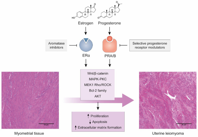
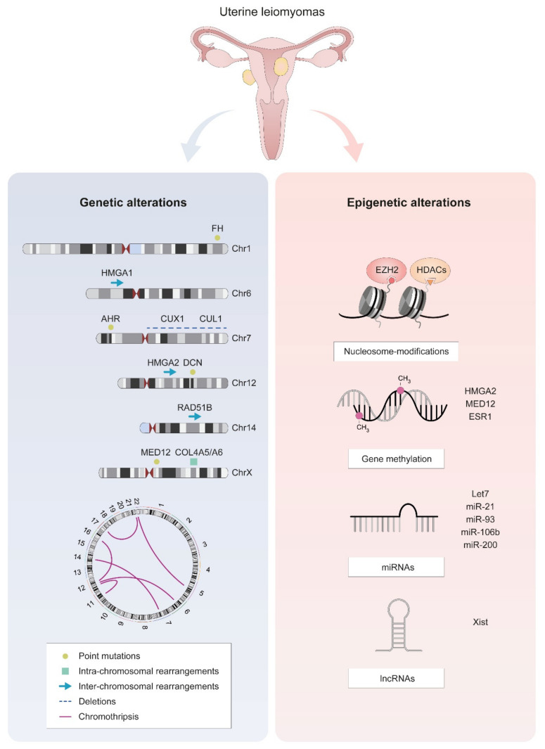
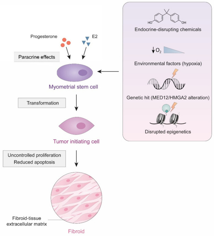
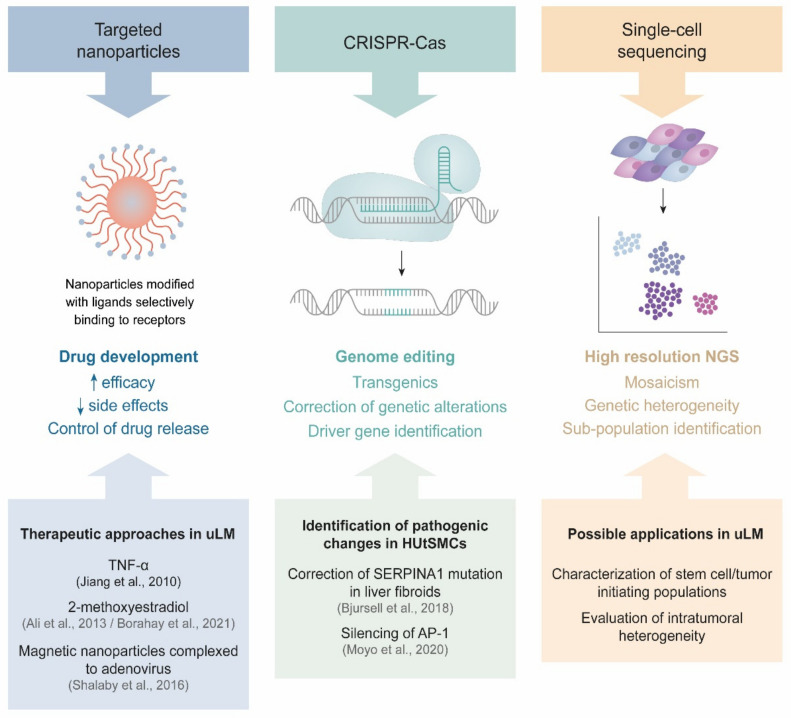
Uterine leiomyomas represent the most common benign gynecologic tumor. These hormone-dependent smooth-muscle formations occur with an estimated prevalence of ~70% among women of reproductive age and cause symptoms including pain, abnormal uterine bleeding, infertility, and recurrent abortion. Despite the prevalence and public health impact of uterine leiomyomas, available treatments remain limited. Among the potential causes of leiomyomas, early hormonal exposure during periods of development may result in developmental reprogramming via epigenetic changes that persist in adulthood, leading to disease onset or progression. Recent developments in unbiased high-throughput sequencing technology enable powerful approaches to detect driver mutations, yielding new insights into the genomic instability of leiomyomas. Current data also suggest that each leiomyoma originates from the clonal expansion of a single transformed somatic stem cell of the myometrium. In this review, we propose an integrated cellular and molecular view of the origins of leiomyomas, as well as paradigm-shifting studies that will lead to better understanding and the future development of non-surgical treatments for these highly frequent tumors.

Keywords: biomarkers; genetics/epigenetics; steroid hormones; targetable pathways; tumor bulk/single-cells; tumor-initiating cell; uterine leiomyoma.


Conflict of interest statement

C.S. is the Founder and Head of the Scientific Advisory Board of Igenomix. The remaining authors declare no conflict of interest.

Figures

Figure 1
Figure 1
Hormonal action on myometrial tissue as a cause of uLM. Images of H&E-stained tissues from our group’s archives. Scale bars represent 100 µm.
Figure 2
Figure 2
The most frequent molecular alterations in uLM cells. Genetic alterations are represented on ideograms of the most frequently affected chromosomes, along with a scheme of HMGA1/2-RAD51B and a circos plot representing chromothripsis. Epigenetic alterations include disrupted nucleosome modification, gene methylation, and altered expression of non-coding RNAs.
Figure 3
Figure 3
Cellular origin of uLM. Specific environmental and genetic changes may cause the transformation of myometrial stem cells into tumor initiating cells, that, through uncontrolled proliferation and reduced apoptosis, may eventually give rise to a uLM tumor.
Figure 4
Figure 4
New techniques in uLM research. Each technique and its features are displayed, with relevant publications or future directions in uLM research.

References

    1. Baird D., Dunson D., Hill M., Cousins D., Schectman J. High cumulative incidence of uterine leiomyoma in black and white women: Ultrasound evidence. Am. J. Obstet. Gynecol. 2003;188:100–107. doi: 10.1067/mob.2003.99. - DOI - PubMed
    1. Stewart E.A., Cookson C.L., Gandolfo R.A., Schulze-Rath R. Epidemiology of uterine fibroids: A systematic review. BJOG. 2017;124:1501–1512. doi: 10.1111/1471-0528.14640. - DOI - PubMed
    1. Marsh E.E., Ekpo G.E., Cardozo E.R., Brocks M., Dune T., Cohen L.S. Racial differences in fibroid prevalence and ultrasound findings in asymptomatic young women (18–30 years old): A pilot study. Fertil. Steril. 2013;99:1951–1957. doi: 10.1016/j.fertnstert.2013.02.017. - DOI - PMC - PubMed
    1. Marsh E.E., Al-Hendy A., Kappus D., Galitsky A., Stewart E.A., Kerolous M. Burden, Prevalence, and Treatment of Uterine Fibroids: A Survey of U.S. Women. J. Women’s Health. 2018;27:1359–1367. doi: 10.1089/jwh.2018.7076. - DOI - PMC - PubMed
    1. Spies J.B., Myers E.R., Worthington-Kirsch R., Mulgund J., Goodwin S., Mauro M. The FIBROID Registry: Symptom and quality-of-life status 1 year after therapy. Obstet. Gynecol. 2005;106:1309–1318. doi: 10.1097/01.AOG.0000188386.53878.49. - DOI - PubMed