Review
. 2021 Aug 8;22(16):8527.
doi: 10.3390/ijms22168527.

The Contribution of Autophagy and LncRNAs to MYC-Driven Gene Regulatory Networks in Cancers

Affiliations
Review

The Contribution of Autophagy and LncRNAs to MYC-Driven Gene Regulatory Networks in Cancers

Leila Jahangiri et al. Int J Mol Sci. .

Abstract

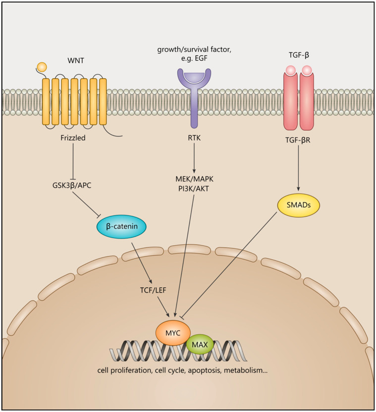
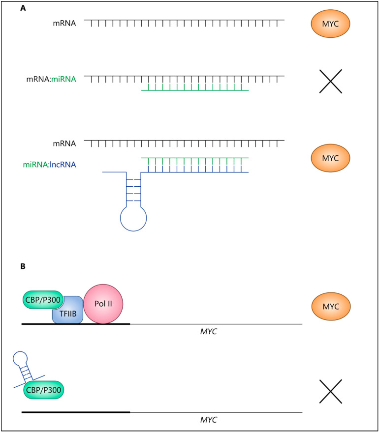
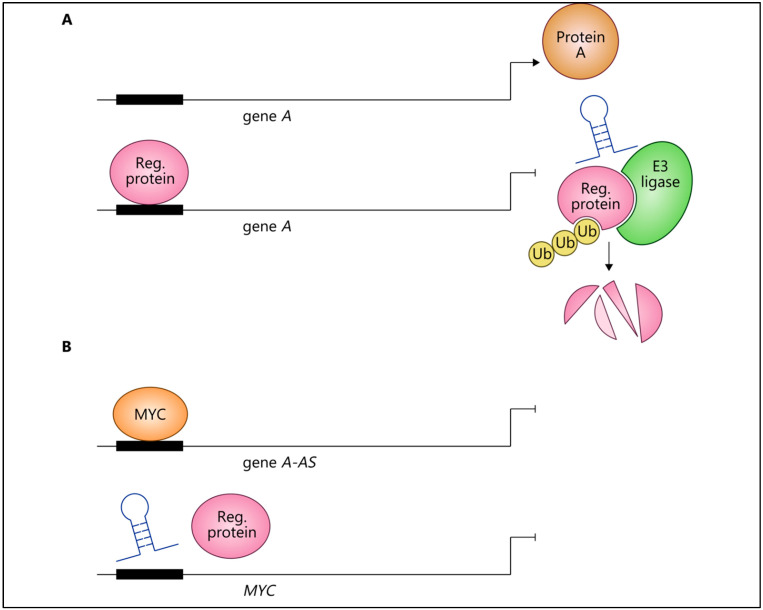
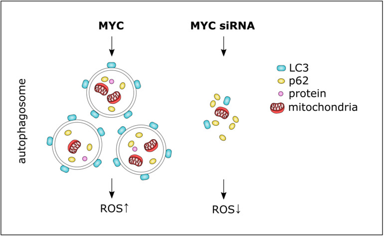
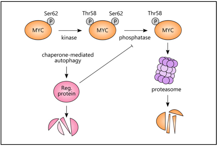
MYC is a target of the Wnt signalling pathway and governs numerous cellular and developmental programmes hijacked in cancers. The amplification of MYC is a frequently occurring genetic alteration in cancer genomes, and this transcription factor is implicated in metabolic reprogramming, cell death, and angiogenesis in cancers. In this review, we analyse MYC gene networks in solid cancers. We investigate the interaction of MYC with long non-coding RNAs (lncRNAs). Furthermore, we investigate the role of MYC regulatory networks in inducing changes to cellular processes, including autophagy and mitophagy. Finally, we review the interaction and mutual regulation between MYC and lncRNAs, and autophagic processes and analyse these networks as unexplored areas of targeting and manipulation for therapeutic gain in MYC-driven malignancies.

Keywords: MYC; autophagy; gene regulatory networks (GRNs); lncRNAs.


Conflict of interest statement

The authors declare no conflict of interest.

Figures

Figure 1
Figure 1
The effects of three signalling pathways converging on MYC activation or repression. A Wnt ligand binds to its receptor frizzled, preventing the phosphorylation of β-catenin by GSK3β and its subsequent degradation. β-catenin accumulates in the nucleus and, in association with T-cell factor/ lymphoid enhancer factor (TCF/LEF), activates Wnt signalling target genes, including MYC. The positive input of growth and survival factors including EGF and its downstream mediators, including MEK/MAPK/PI3K/AKT on MYC activation, while TGF-β signalling via SMADs can suppress MYC activity. MYC can form a heterodimer with MAX and can govern cell proliferation, apoptosis, and metabolism.
Figure 2
Figure 2
Regulatory loops involving lncRNAs and MYC. (A) A lncRNA (e.g., MALAT1) acting as a competing endogenous RNA by sequestering miRNA (e.g., miR-204), allowing for the production of MYC. (B) The physical interaction between a lncRNA (e.g., linc00261) prevents the recruitment of p300/CBP to the promoter region of MYC, thereby repressing MYC expression.
Figure 3
Figure 3
A negative feedback loop involving MYC, lncRNAs, and ubiquitin ligase. (A) LncRNA (encoded by gene A-AS1, e.g., MTSS1-AS1) upregulated the expression of gene A (e.g., MTSS1 gene) acting as a scaffold between E3 ligase and a regulatory protein (e.g., MZF1), leading to ubiquitination-mediated degradation of the regulatory protein; hence, protein A (e.g., MTSS1) was produced. The regulatory protein (e.g., MZF1) inhibited gene A (e.g., MTSS1 gene) expression by binding its promoter. (BMYC inhibited the lncRNA (e.g., MTSS1-AS1) by binding its initiator elements (gene A-AS, e.g., MTSS1-AS1). In turn, the lncRNA (encoded by gene A-AS, e.g., MTSS1-AS1) inhibited MYC expression by impairing its regulatory protein-mediated transcriptional activation. Reg. = regulatory.
Figure 4
Figure 4
MYC and autophagy. MYC was involved in autophagosome formation whereby siRNA-mediated knockdown of MYC inhibited autophagy, resulting in the accumulation of the autophagy substrate p62. MYC is known to trigger ROS accumulation.
Figure 5
Figure 5
CMA and MYC protein stability. A phosphatase removes phosphoserine 62 from MYC, rendering it unstable and susceptible to proteasomal degradation. This phosphatase may be suppressed by a regulatory protein (e.g., CIP2A) that can, in turn, be degraded by CMA-associated processes. Reg. = regulatory.

References

    1. Malynn B.A., de Alboran I.M., O’Hagan R.C., Bronson R., Davidson L., DePinho R.A., Alt F.W. N-myc can functionally replace c-myc in murine development, cellular growth, and differentiation. Genes Dev. 2000;14:1390–1399. - PMC - PubMed
    1. Kohl N.E., Kanda N., Schreck R.R., Bruns G., Latt S.A., Gilbert F., Alt F.W. Transposition and amplification of oncogene-related sequences in human neuroblastomas. Cell. 1983;35:359–367. doi: 10.1016/0092-8674(83)90169-1. - DOI - PubMed
    1. Amati B., Littlewood T.D., Evan G.I., Land H. The c-Myc protein induces cell cycle progression and apoptosis through dimerization with Max. EMBO J. 1993;12:5083–5087. doi: 10.1002/j.1460-2075.1993.tb06202.x. - DOI - PMC - PubMed
    1. Dang C.V. MYC on the path to cancer. Cell. 2012;149:22–35. doi: 10.1016/j.cell.2012.03.003. - DOI - PMC - PubMed
    1. Soucek L., Evan G.I. The ups and downs of Myc biology. Curr. Opin. Genet. Dev. 2010;20:91–95. doi: 10.1016/j.gde.2009.11.001. - DOI - PMC - PubMed

MeSH terms

LinkOut - more resources