. 2021 Aug 9;22(16):8547.
doi: 10.3390/ijms22168547.

Chitin-Derived AVR-48 Prevents Experimental Bronchopulmonary Dysplasia (BPD) and BPD-Associated Pulmonary Hypertension in Newborn Mice

Affiliations

Chitin-Derived AVR-48 Prevents Experimental Bronchopulmonary Dysplasia (BPD) and BPD-Associated Pulmonary Hypertension in Newborn Mice

Pragnya Das et al. Int J Mol Sci. .

Abstract

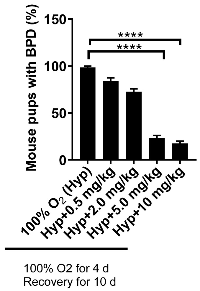
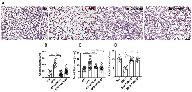
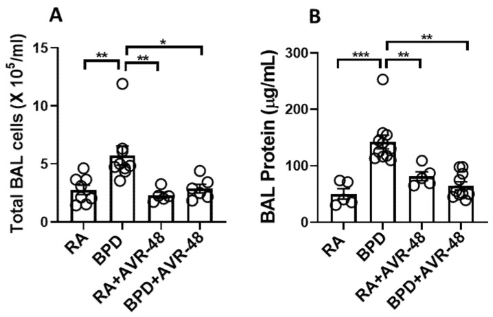
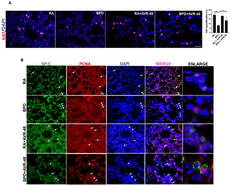
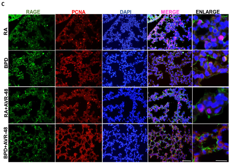
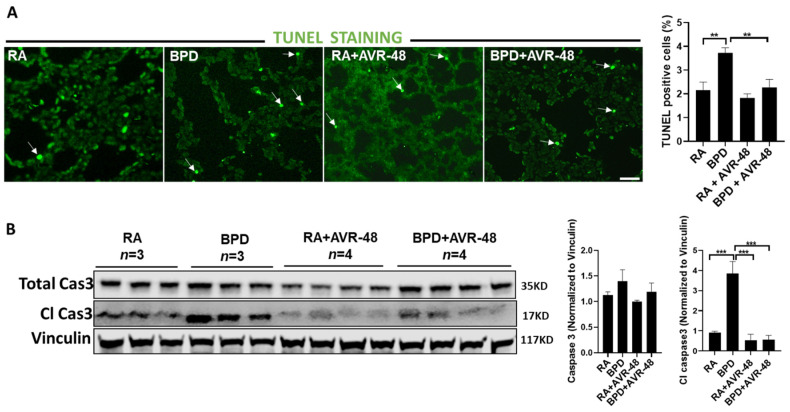
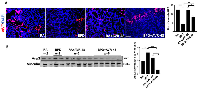
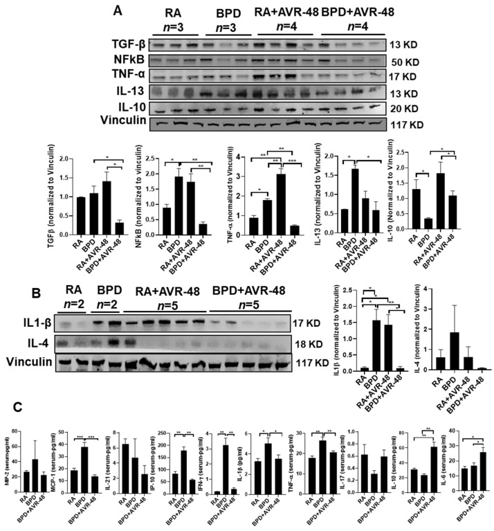
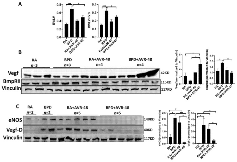
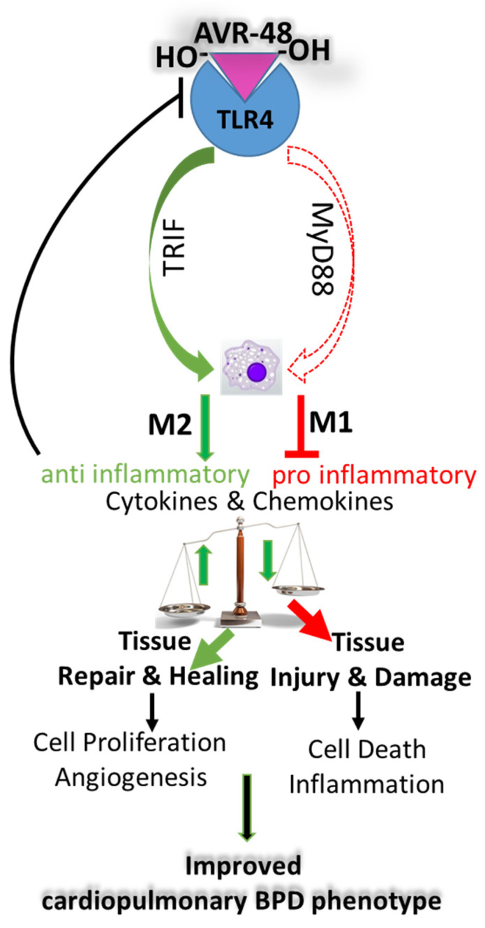
Bronchopulmonary dysplasia (BPD) is the most common complication of prematurity and a key contributor to the large health care burden associated with prematurity, longer hospital stays, higher hospital costs, and frequent re-hospitalizations of affected patients through the first year of life and increased resource utilization throughout childhood. This disease is associated with abnormal pulmonary function that may lead to BPD-associated pulmonary hypertension (PH), a major contributor to neonatal mortality and morbidity. In the absence of any definitive treatment options, this life-threatening disease is associated with high resource utilization during and after neonatal intensive care unit (NICU) stay. The goal of this study was to test the safety and efficacy of a small molecule derivative of chitin, AVR-48, as prophylactic therapy for preventing experimental BPD in a mouse model. Two doses of AVR-48 were delivered either intranasally (0.11 mg/kg), intraperitoneally (10 mg/kg), or intravenously (IV) (10 mg/kg) to newborn mouse pups on postnatal day (P)2 and P4. The outcomes were assessed by measuring total inflammatory cells in the broncho-alveolar lavage fluid (BALF), chord length, septal thickness, and radial alveolar counts of the alveoli, Fulton's Index (for PH), cell proliferation and cell death by immunostaining, and markers of inflammation by Western blotting and ELISA. The bioavailability and safety of the drug were assessed by pharmacokinetic and toxicity studies in both neonatal mice and rat pups (P3-P5). Following AVR-48 treatment, alveolar simplification was improved, as evident from chord length, septal thickness, and radial alveolar counts; total inflammatory cells were decreased in the BALF; Fulton's Index was decreased and lung inflammation and cell death were decreased, while angiogenesis and cell proliferation were increased. AVR-48 was found to be safe and the no-observed-adverse-effect level (NOAEL) in rat pups was determined to be 100 mg/kg when delivered via IV dosing with a 20-fold safety margin. With no reported toxicity and with a shorter half-life, AVR-48 is able to reverse the worsening cardiopulmonary phenotype of experimental BPD and BPD-PH, compared to controls, thus positioning it as a future drug candidate.

Keywords: AVR-48; BPD-associated pulmonary hypertension; bronchopulmonary dysplasia; chitohexaose; inflammation.


Conflict of interest statement

V.B. is on the Scientific Advisory Board of AyuVis Research Inc. for development of BPD drugs.

Figures

Figure 1
Figure 1
Dose response study of AVR-48 at different doses of 0.5, 2.0, 5.0, and 10 mg/kg, given IP. **** p < 0.0001. n = 5–7 mice per group. 10 mg/kg (IP) was selected as the most efficacious dose. IP: intraperitoneal; Hyp: hyperoxia; BPD: bronchopulmonary dysplasia.
Figure 2
Figure 2
AVR-48 improves lung morphology. (A) Representative H&E stained lung paraffin sections showing histological changes after AVR-48 treatment. (B) Chord length (which measures the average free distance in the air spaces) is increased in the BPD group and normalizes after AVR-48 treatment. (C) The alveolar septal thickness is decreased and (D) the radial alveolar count (which measures the number of alveoli) is also improved after AVR-48 treatment. *** p < 0.001, n = 3–8; RA: room air; BPD: bronchopulmonary dysplasia. Scale bar 100 µm.
Figure 3
Figure 3
AVR-48 decreases inflammation and vascular leak. (A) Total inflammatory cells in the BAL fluid in the BPD group is significantly decreased after AVR-48 treatment. (B) Total protein in the BAL fluid in the BPD group is significantly decreased after AVR-48 treatment. * p < 0.05; ** p < 0.01; *** p < 0.001, n = 3–8; RA: room air; BAL: bronchoalveolar lavage; BPD: bronchopulmonary dysplasia.
Figure 4
Figure 4
AVR-48 improves cell proliferation (A) AVR-48 treatment in the BPD group increases cell proliferation (as shown by Ki67 staining) and the right panel shows quantification for Ki67. (B) Co-localization of SP-C (marker for Type II AECs) with PCNA. White arrows point to the respective cells that are proliferating. Extreme right panel shows higher magnification of proliferating Type II AECs positive for SP-C (cytoplasmic green) and PCNA (nuclear red). (C) Co-localization of RAGE (marker for Type I AECs) with PCNA. Extreme right panel shows higher magnification of proliferating Type I cells positive for RAGE (cytoplasmic green) and PCNA (nuclear red). ** p < 0.01; *** p < 0.001; Scale bar 100 µm. RA: room air; BPD: bronchopulmonary dysplasia; SP: surfactant protein; AECs: alveolar epithelial cells; PCNA: proliferating cell nuclear antigen; RAGE: receptor for advanced glycation end products.
Figure 4
Figure 4
AVR-48 improves cell proliferation (A) AVR-48 treatment in the BPD group increases cell proliferation (as shown by Ki67 staining) and the right panel shows quantification for Ki67. (B) Co-localization of SP-C (marker for Type II AECs) with PCNA. White arrows point to the respective cells that are proliferating. Extreme right panel shows higher magnification of proliferating Type II AECs positive for SP-C (cytoplasmic green) and PCNA (nuclear red). (C) Co-localization of RAGE (marker for Type I AECs) with PCNA. Extreme right panel shows higher magnification of proliferating Type I cells positive for RAGE (cytoplasmic green) and PCNA (nuclear red). ** p < 0.01; *** p < 0.001; Scale bar 100 µm. RA: room air; BPD: bronchopulmonary dysplasia; SP: surfactant protein; AECs: alveolar epithelial cells; PCNA: proliferating cell nuclear antigen; RAGE: receptor for advanced glycation end products.
Figure 5
Figure 5
AVR-48 decreases cell death: TUNEL staining (white arrows point to TUNEL positive cells) (A) and Western blotting of total caspase 3 and cleaved caspase 3 (B) shows decrease in cell death and apoptosis after treatment with AVR-48. Right panel shows quantification of TUNEL positive cells (top) and densitometric quantification of total caspase 3 and cleaved caspase 3 (bottom). n = 3–4. ** p < 0.01; ***p < 0.001; Scale bar 100 µm. RA: room air; BPD: bronchopulmonary dysplasia; Cl Cas: cleaved caspase.
Figure 6
Figure 6
AVR-48 promotes vascular development. (A) Representative immunofluorescent lung sections showing vascular development. vWF, a marker for blood vessels, is severely disrupted in BPD, while after treatment with AVR-48 there is significant improvement. (B) Representative Western blotting showing Ang2 is restored after treatment with AVR-48, in the BPD group. The top right panel shows quantification of the number of blood vessels while the bottom right panel shows densitometric quantification for Ang2. Scale bar 100 µm; * p < 0.05; ** p < 0.01; *** p < 0.001; vWF: von Willebrand factor; Ang2: angiopoietin 2; RA: room air; BPD: bronchopulmonary dysplasia; HPF: high power field; n = 3–5.
Figure 7
Figure 7
AVR-48 suppresses inflammation. (A,B) Representative Western blot showing decrease of pro-inflammatory cytokines (TGFβ, NFkB, TNFα, IL-13, IL-1β, and IL-4) and increase of IL-10 in the lungs after treatment with AVR-48, as compared to the BPD group. The increased inflammation seen in the RA+AVR-48 treated group may be due to the natural defense adaptive mechanism. Vinculin is the loading control. The panel below the gel shows densitometric quantification of the proteins. n = 5. (C) ELISA showing the expression of selected cytokines in the blood serum of treated BPD group as compared to untreated BPD controls. The RA+AVR-48 group was not included for this assay. Although most of the pro-inflammatory cytokines and chemokines show a decrease after treatment, there was no change in MIP-2, IL-21, or IL-17. * p < 0.05; ** p < 0.01; *** p < 0.001, n = 4–5; RA: room air; BPD: bronchopulmonary dysplasia; TGFβ: transforming growth factor beta; MCP-1: monocyte chemoattractant protein 1; MIP-2: macrophage inflammatory protein 2; NfkB: nuclear factor kappa B; TNFα: tumor necrosis factor alpha; IFNγ: interferon gamma; IL: interleukin.
Figure 8
Figure 8
AVR-48 protects against BPD-PH. (A) The RV/LV ratio and Fulton’s Index (RV/LV+IVS) is improved after AVR-48 treatment in the BPD group. (B) Representative Western blot showing an increased expression of Vegf in the BPD+AVR-48 treated group as compared to BPD group. (C) eNOS, BmpRII and VegfD, which are increased in BPD, are noticeably decreased after treatment with AVR-48. Vinculin is the loading control. As the same samples were used for Figure 7A,B and Figure 8B,C, the same vinculin has been shown for both images as the loading control. n = 4–5. BPD-PH: bronchopulmonary dysplasia-associated pulmonary hypertension; RV: right ventricle; LV: left ventricle; IVS: interventricular septum; RA: room air; BPD: bronchopulmonary dysplasia; Vegf: vascular endothelial growth factor; eNOS: endothelial nitric oxide synthase; BmpRII: bone morphogenetic protein receptor 2. * p < 0.05; ** p < 0.01; *** p < 0.001.
Figure 9
Figure 9
Proposed mechanism of action of AVR-48 in neonatal lungs. AVR-48 after binding to TLR4 triggers the TRIF pathway to activate the M2 macrophages via the alternate pathway to produce IL-10, which in turn negatively regulates TLR4 to downregulate the MyD88 pathway to decrease the synthesis of a myriad of pro-inflammatory cytokines and chemokines by suppressing the M1 macrophages that are produced via activation of the classical pathway during BPD. This combinatorial effect results in decreasing tissue injury and increasing tissue repair and healing by maintaining a balance between M2 and M1 macrophages toward a favorable outcome with overall improvement of the BPD cardiopulmonary phenotype.

References

    1. Bhandari V. Bronchopulmonary Dysplasia. Springer; Berlin/Heidelberg, Germany: 2016.
    1. Bhandari A., Bhandari V. Pitfalls, problems, and progress in bronchopulmonary dysplasia. Pediatrics. 2009;123:1562–1573. doi: 10.1542/peds.2008-1962. - DOI - PubMed
    1. Bhandari V. Drug therapy trials for the prevention of bronchopulmonary dysplasia: Current and future targets. Front. Pediatr. 2014;2:76. doi: 10.3389/fped.2014.00076. - DOI - PMC - PubMed
    1. Sahni M., Bhandari V. Recent advances in understanding and management of bronchopulmonary dysplasia. F1000Research. 2020;9:703. doi: 10.12688/f1000research.25338.1. - DOI - PMC - PubMed
    1. Lui K., Lee S.K., Kusuda S., Adams M., Vento M., Reichman B., Darlow B.A., Lehtonen L., Modi N., Norman M., et al. Trends in Outcomes for Neonates Born Very Preterm and Very Low Birth Weight in 11 High-Income Countries. J. Pediatr. 2019;215:32–40.e14. doi: 10.1016/j.jpeds.2019.08.020. - DOI - PubMed

MeSH terms