Review
. 2021 Aug 10;22(16):8580.
doi: 10.3390/ijms22168580.

Platelet-Derived Extracellular Vesicles for Regenerative Medicine

Affiliations
Review

Platelet-Derived Extracellular Vesicles for Regenerative Medicine

Miquel Antich-Rosselló et al. Int J Mol Sci. .

Abstract

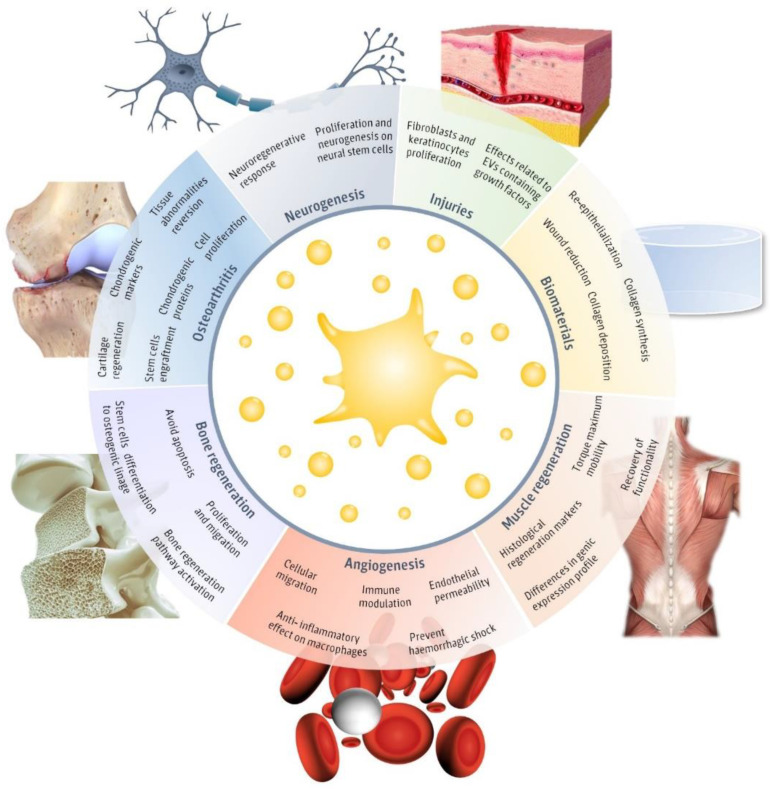
Extracellular vesicles (EVs) present a great potential for the development of new treatments in the biomedical field. To be used as therapeutics, many different sources have been used for EVs obtention, while only a few studies have addressed the use of platelet-derived EVs (pEVs). In fact, pEVs have been shown to intervene in different healing responses, thus some studies have evaluated their regenerative capability in wound healing or hemorrhagic shock. Even more, pEVs have proven to induce cellular differentiation, enhancing musculoskeletal or neural regeneration. However, the obtention and characterization of pEVs is widely heterogeneous and differs from the recommendations of the International Society for Extracellular Vesicles. Therefore, in this review, we aim to present the main advances in the therapeutical use of pEVs in the regenerative medicine field while highlighting the isolation and characterization steps followed. The main goal of this review is to portray the studies performed in order to enhance the translation of the pEVs research into feasible therapeutical applications.

Keywords: exosomes; extracellular vesicles; platelets; regenerative medicine.


Conflict of interest statement

The authors declare no conflict of interest.

Figures

Figure 1
Figure 1
Regenerative applications of platelet-derived extracellular vesicles (pEVs). Main regenerative effects reported for pEVs in regenerative fields, including injuries [30,31,32,33,34,35,36], biomaterials [30,31], neurogenesis [37,38], muscle regeneration [39], angiogenesis [37,38,40,41,42], bone regeneration [25,26,43,44,45] and osteoarthritis [46,47,48,49] and the major reported therapeutical effects. This figure was created using Freepik images.
Figure 2
Figure 2
pEVs isolation methods reported for regenerative approaches. (a) Diagram shows the proportion and the number of reports that used centrifugation at low relative centrifugal force (RCF; lower than 80,000× g), centrifugation at high RCF (higher than 80,000× g), filtration, size exclusion chromatography, other methods or did not report the isolation method used. If two different methods or a combination of methods was used, both groups are represented on the diagram. (b) Diagram represents the platelet source used for pEVs isolation, whether it was platelet rich plasma (PRP), activated PRP or other platelet sources. (c) Diagram shows the storage conditions of the isolated pEVs before use. Temperature conditions at −80 °C, −20 °C, others or not reported conditions are also represented on the figure. A total of 22 articles were reviewed to obtain this data.
Figure 3
Figure 3
Characterization reported for pEVs used in regenerative applications. (a) Number of articles and the percentage of them which report physical characterization (include nanoparticle tracking analysis, electron microscopy, flow cytometry or dynamic light scattering), and pEV marker detection (include immunolabelling through western blot, flow cytometry or electron microscopy); (b) Number of articles and the percentage of them which report the different EV markers: pEV membrane marker (CD9, CD61, CD63 or CD81), platelet source marker (CD31, CD41 or CD42), pEV cytosolic markers (ALIX, TSG101, HSP90 or HSP101) and non-EV structures (APOA1, APOB100 or calnexin). A total of 22 articles were reviewed to obtain this data.
Figure 4
Figure 4
Physical and biochemical characteristics of pEVs depending on their origin.

References

    1. Théry C., Witwer K.W., Aikawa E., Alcaraz M.J., Anderson J.D., Andriantsitohaina R., Antoniou A., Arab T., Archer F., Atkin-Smith G.K., et al. Minimal information for studies of extracellular vesicles 2018 (MISEV2018): A position statement of the International Society for Extracellular Vesicles and update of the MISEV2014 guidelines. J. Extracell. Vesicles. 2018;7:1535750. doi: 10.1080/20013078.2018.1535750. - DOI - PMC - PubMed
    1. Colombo M., Raposo G., Théry C. Biogenesis, secretion, and intercellular interactions of exosomes and other extracellular vesicles. Annu. Rev. Cell Dev. Biol. 2014;30:255–289. doi: 10.1146/annurev-cellbio-101512-122326. - DOI - PubMed
    1. Doyle L.M., Wang M.Z. Overview of Extracellular Vesicles, Their Origin, Composition, Purpose, and Methods for Exosome Isolation and Analysis. Cells. 2019;8:727. doi: 10.3390/cells8070727. - DOI - PMC - PubMed
    1. Van Niel G., D’Angelo G., Raposo G. Shedding light on the cell biology of extracellular vesicles. Nat. Rev. Mol. Cell Biol. 2018;19:213–228. doi: 10.1038/nrm.2017.125. - DOI - PubMed
    1. Mathieu M., Martin-Jaular L., Lavieu G., Théry C. Specificities of secretion and uptake of exosomes and other extracellular vesicles for cell-to-cell communication. Nat. Cell Biol. 2019;21:9–17. doi: 10.1038/s41556-018-0250-9. - DOI - PubMed

MeSH terms

LinkOut - more resources