Review
. 2021 Aug 10;22(16):8590.
doi: 10.3390/ijms22168590.

Insulin Signal Transduction Perturbations in Insulin Resistance

Affiliations
Review

Insulin Signal Transduction Perturbations in Insulin Resistance

Mariyam Khalid et al. Int J Mol Sci. .

Abstract

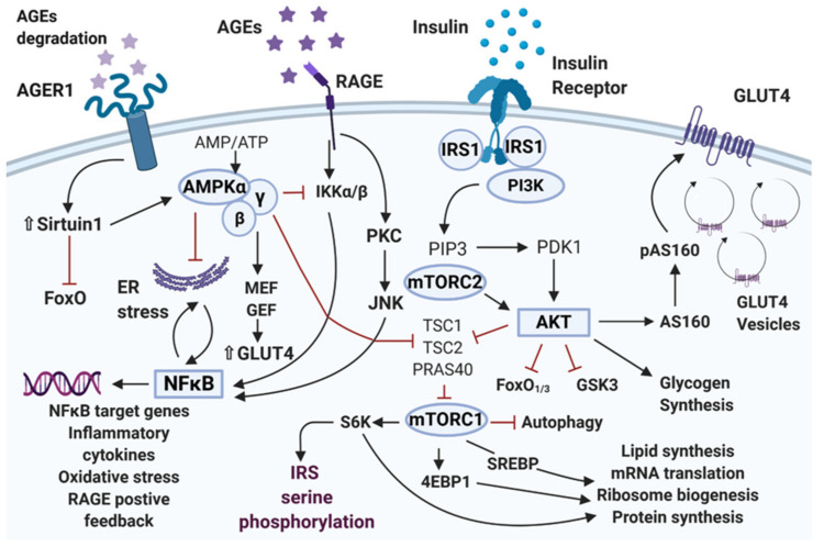
Type 2 diabetes mellitus is a widespread medical condition, characterized by high blood glucose and inadequate insulin action, which leads to insulin resistance. Insulin resistance in insulin-responsive tissues precedes the onset of pancreatic β-cell dysfunction. Multiple molecular and pathophysiological mechanisms are involved in insulin resistance. Insulin resistance is a consequence of a complex combination of metabolic disorders, lipotoxicity, glucotoxicity, and inflammation. There is ample evidence linking different mechanistic approaches as the cause of insulin resistance, but no central mechanism is yet described as an underlying reason behind this condition. This review combines and interlinks the defects in the insulin signal transduction pathway of the insulin resistance state with special emphasis on the AGE-RAGE-NF-κB axis. Here, we describe important factors that play a crucial role in the pathogenesis of insulin resistance to provide directionality for the events. The interplay of inflammation and oxidative stress that leads to β-cell decline through the IAPP-RAGE induced β-cell toxicity is also addressed. Overall, by generating a comprehensive overview of the plethora of mechanisms involved in insulin resistance, we focus on the establishment of unifying mechanisms to provide new insights for the future interventions of type 2 diabetes mellitus.

Keywords: hyperglycemia; insulin resistance; insulin signaling; pancreatic beta cells; type 2 diabetes mellitus.


Conflict of interest statement

The authors declare that there are no conflicts of interest.

Figures

Figure 1
Figure 1
Various pathways involved in dysregulation of insulin signaling. Upon insulin binding, insulin receptor gets autophosphorylated. This recruits different substrate adaptors for the signal transduction. The tyrosine phosphorylated IRS1 recruits PI3K, which phosphorylates the serine/threonine residue of protein kinase B (Akt). Akt regulates the translocation of glucose transporter GLUT4 to the cell surface through phosphorylation of the GTPase-activating protein (AS160). Akt promotes glycogen synthesis through inhibition of GSK3 activity and induces protein synthesis via activation of mTOR and downstream elements. Akt phosphorylates and directly inhibits FoxO transcription factors, which inhibits autophagy. The hyperglycemia-induced production of AGE and binding with their receptor RAGE impairs insulin signal transduction by triggering a range of signaling pathways, including JNK, NF-κB, and activation of PKC. The sustained accumulation of AGE depletes the expression of anti-AGE cell surface receptor AGER1, which is responsible for inhibiting the deleterious effects of AGE by competitively interfering with its binding to RAGE. AGER1 along with Sirtuin1 promotes AMPK phosphorylation and activation, which induces GLUT4 gene expression through activation of MEF, GEF transcription factors. The AGE-RAGE induced activation of PKC, NF-κB mediated inflammation, and oxidative stress promotes the serine phosphorylation of IRS, inhibits its action, and induces insulin resistance. AGE: Advanced glycation end products; AGER1: AGE receptor 1 encoded by DDOST gene; AMPK: AMP-activated protein kinase; AS160: Akt substrate of 160 kDa; FoxO: Forkhead family of transcription factors; GLUT4: Glucose transporter protein 4; GEF: GLUT4 enhancer factor; GSK3: Glycogen synthase kinase 3; IKK: IκB kinase; IRS: Insulin receptor substrate1; MEF: Myocyte enhancer factor; mTOR: Mammalian target of rapamycin NF-κB: Nuclear factor κB; PDK1: Phosphoinositide-dependent kinase 1; PI3K: Phosphoinositide 3-kinase; PIP3: Phosphatidylinositol (3,4,5)-trisphosphate; PKC: Protein kinase C; RAGE: Receptor for advanced glycosylation end products; S6K: Ribosomal protein S6 kinase; SREBP: Sterol regulatory element-binding proteins; JNK: c-Jun N-terminal kinase.

References

    1. Barnett D.M., Krall L.P. Joslin’s Diabetes Mellitus. Lippincott Williams & Wilkins; Philadelphia, PA, USA: 2005. The history of diabetes; pp. 1–17.
    1. Kang S., Tsai L.T.Y., Rosen E.D. Nuclear Mechanisms of Insulin Resistance. Trends Cell Biol. 2016;26:341–351. doi: 10.1016/j.tcb.2016.01.002. - DOI - PMC - PubMed
    1. Petersen M.C., Shulman G.I. Mechanisms of Insulin Action and Insulin Resistance. Physiol. Rev. 2018;98:2133–2223. doi: 10.1152/physrev.00063.2017. - DOI - PMC - PubMed
    1. Mlinar B., Marc J., Janež A., Pfeifer M. Molecular Mechanisms of Insulin Resistance and Associated Diseases. Clin. Chim. Acta. 2007;375:20–35. doi: 10.1016/j.cca.2006.07.005. - DOI - PubMed
    1. Mezza T., Cinti F., Cefalo C.M.A., Pontecorvi A., Kulkarni R.N., Giaccari A. β-Cell Fate in Human Insulin Resistance and Type 2 Diabetes: A Perspective on Islet Plasticity. Diabetes. 2019;68:1121–1129. doi: 10.2337/db18-0856. - DOI - PMC - PubMed

MeSH terms