. 2021 Aug 10;22(16):8596.
doi: 10.3390/ijms22168596.

Cathepsin L, a Target of Hypoxia-Inducible Factor-1-α, Is Involved in Melanosome Degradation in Melanocytes

Affiliations

Cathepsin L, a Target of Hypoxia-Inducible Factor-1-α, Is Involved in Melanosome Degradation in Melanocytes

Ji Young Kim et al. Int J Mol Sci. .

Abstract

Hypoxic conditions induce the activation of hypoxia-inducible factor-1α (HIF-1α) to restore the supply of oxygen to tissues and cells. Activated HIF-1α translocates into the nucleus and binds to hypoxia response elements to promote the transcription of target genes. Cathepsin L (CTSL) is a lysosomal protease that degrades cellular proteins via the endolysosomal pathway. In this study, we attempted to determine if CTSL is a hypoxia responsive target gene of HIF-1α, and decipher its role in melanocytes in association with the autophagic pathway. The results of our luciferase reporter assay showed that the expression of CTSL is transcriptionally activated through the binding of HIF1-α at its promoter. Under autophagy-inducing starvation conditions, HIF-1α and CTSL expression is highly upregulated in melan-a cells. The mature form of CTSL is closely involved in melanosome degradation through lysosomal activity upon autophagosome-lysosome fusion. The inhibition of conversion of pro-CTSL to mature CTSL leads to the accumulation of gp100 and tyrosinase in addition to microtubule-associated protein 1 light chain 3 (LC3) II, due to decreased lysosomal activity in the autophagic pathway. In conclusion, we have identified that CTSL, a novel target of HIF-1α, participates in melanosome degradation in melanocytes through lysosomal activity during autophagosome-lysosome fusion.

Keywords: Cathepsin L; autophagy; hypoxia-inducible factor-1-alpha; melanosome.


Conflict of interest statement

The authors declare no conflict of interest.

Figures

Figure 1
Figure 1
Hypoxic condition enhanced HIF-1α and CTSL activation. (a) Western blotting analysis of cytosolic and nuclear fractions of HIF-1α under normoxic or hypoxic conditions. mRNA (b) and protein (c) levels of CTSL were assessed in melan-a cells in hypoxia. (d) HREs in CTSL promoter were predicted. (e) Cells were transfected with luciferase reporter vectors, pGL3-Ctsl/HRE or pGL3-Ctsl/mutHRE, and luciferase activity was determined under hypoxic conditions for 4 h. (f) ChIP-qPCR analysis for HIF1-α occupancy on CTSL promoter in melan-a cells in normoxa or hypoxia for 4 h. Quantification of enrichment was represented as fold-enrichment relative to IgG. Results are expressed as mean ± S.D of more than three independent experiments. * p < 0.05. Student’s t-test.
Figure 2
Figure 2
HIF-1α and CTSL induction was enhanced by starvation condition in melan-a cells. (a) mRNA levels of HIF-1α and CTSL were evaluated under starvation control. Moreover, (b) the protein level of CTSL was examined. Melan-a cells were pretreated with CT for 1 h and (c) mRNA levels of HIF-1α and CTSL were determined under starvation for 24 h. (d) Protein level of CTSL was also confirmed. Results are expressed as mean ± S.D of more than three independent experiments. * p < 0.05. Student’s t-test.
Figure 3
Figure 3
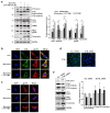
CTSL is associated with melanosome degradation through autophagy in melan-a cells. (a) Protein levels of p62, LC3, PMEL/gp100, and tyrosinase were examined under starvation conditions. (b) Melan-a cells were treated with Z-FF-FMK for 1 h and protein levels of CTSL, LC3, PMEL/gp100, and tyrosinase were evaluated. (c) After Z-FF-FMK pre-treatment and starvation for 8 h, tandem fluorescent-tagged LC3 (mRFP-GFP-LC3) was observed by confocal microscopy. Yellow (autophagosome) and red (autolysosome). Scale bar: 10 μm. Results are expressed as mean ± S.D of more than three independent experiments. * p < 0.05. Student’s t-test.
Figure 4
Figure 4
Inhibition of CTSL activity led to accumulation of melanosome-related molecules in melan-a cells. (a) After Z-FF-FMK pre-treatment and starvation for 24 h, CTSL, LC3, PMEL/gp100, and tyrosinase protein expressions were determined. (b) CTSL and gp100 expressions were observed by confocal microscopy after Z-FF-FMK pre-treatment and starvation for 4 h. Scale bar: 10 μm. (c) gp100 expression was observed by confocal microscopy after Z-FF-FMK pre-treatment and starvation for 24 h. Scale bar: 10 μm. (d) Confocal image of melan-a cells after scramble (scr) siRNA or CTSL siRNA treatment for 48 h. Scale bar: 20 μm. (e) CTSL, LC3, PMEL/gp100, and tyrosinase protein expressions were evaluated after scramble (scr) siRNA or CTSL siRNA treatment for 48 h. Results are expressed as mean ± S.D of more than three independent experiments. * p < 0.05. Student’s t-test.

References

    1. Semenza G.L. Hypoxia-Inducible Factors in Physiology and Medicine. Cell. 2012;148:399–408. doi: 10.1016/j.cell.2012.01.021. - DOI - PMC - PubMed
    1. Zhou M., Xie Y., Xu S., Xin J., Wang J., Han T., Ting R., Zhang J., An F. Hypoxia-activated nanomedicines for effective cancer therapy. Eur. J. Med. Chem. 2020;195:112274. doi: 10.1016/j.ejmech.2020.112274. - DOI - PubMed
    1. Lee J.-W., Bae S.-H., Jeong J.-W., Kim S.-H., Kim K.-W. Hypoxia-inducible factor (HIF-1)alpha: Its protein stability and biological functions. Exp. Mol. Med. 2004;36:1–12. doi: 10.1038/emm.2004.1. - DOI - PubMed
    1. Wang G.L., Jiang B., Rue E.A., Semenza G.L. Hypoxia-inducible factor 1 is a basic-helix-loop-helix-PAS heterodimer regulated by cellular O2 tension. Proc. Natl. Acad. Sci. USA. 1995;92:5510–5514. doi: 10.1073/pnas.92.12.5510. - DOI - PMC - PubMed
    1. Wenger R., Stiehl D.P., Camenisch G. Integration of oxygen signaling at the consensus HRE. Sci. STKE. 2005;2005:re12. doi: 10.1126/stke.3062005re12. - DOI - PubMed

Substances