. 2021 Aug 12;22(16):8675.
doi: 10.3390/ijms22168675.

Patient-Derived Organoids of Cholangiocarcinoma

Affiliations

Patient-Derived Organoids of Cholangiocarcinoma

Christopher Fabian Maier et al. Int J Mol Sci. .

Abstract

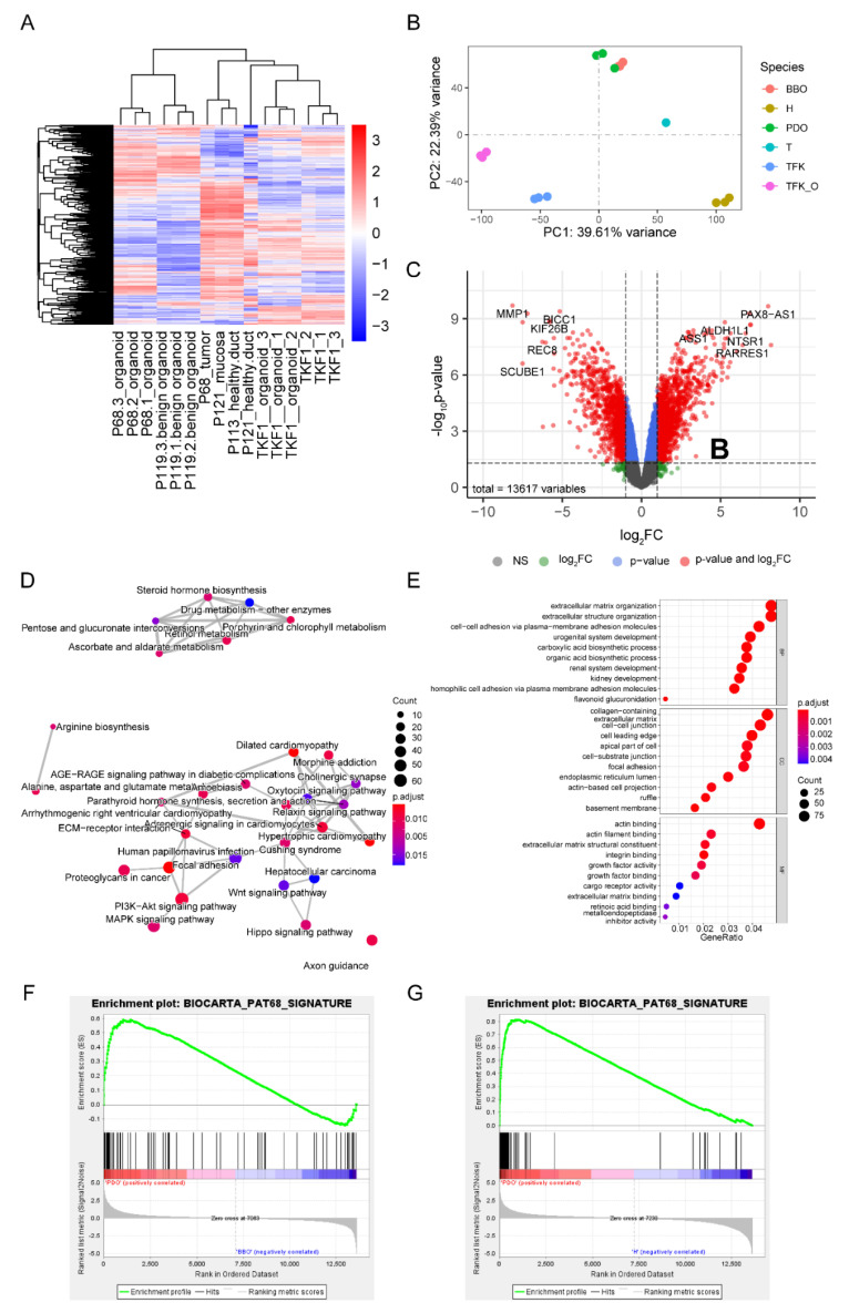
Cholangiocarcinoma (CC) is an aggressive malignancy with an inferior prognosis due to limited systemic treatment options. As preclinical models such as CC cell lines are extremely rare, this manuscript reports a protocol of cholangiocarcinoma patient-derived organoid culture as well as a protocol for the transition of 3D organoid lines to 2D cell lines. Tissue samples of non-cancer bile duct and cholangiocarcinoma were obtained during surgical resection. Organoid lines were generated following a standardized protocol. 2D cell lines were generated from established organoid lines following a novel protocol. Subcutaneous and orthotopic patient-derived xenografts were generated from CC organoid lines, histologically examined, and treated using standard CC protocols. Therapeutic responses of organoids and 2D cell lines were examined using standard CC agents. Next-generation exome and RNA sequencing was performed on primary tumors and CC organoid lines. Patient-derived organoids closely recapitulated the original features of the primary tumors on multiple levels. Treatment experiments demonstrated that patient-derived organoids of cholangiocarcinoma and organoid-derived xenografts can be used for the evaluation of novel treatments and may therefore be used in personalized oncology approaches. In summary, this study establishes cholangiocarcinoma organoids and organoid-derived cell lines, thus expanding translational research resources of cholangiocarcinoma.

Keywords: cholangiocarcinoma; next-generation sequencing; organoids; orthotopic xenograft; patient-derived organoids; precision medicine; response prediction; translational surgical oncology; xenograft model.


Conflict of interest statement

The authors declare no conflict of interest. The funders had no role in the design of the study, in the collection, analyses, or interpretation of data, in the writing of the manuscript, or in the decision to publish the results.

Figures

Figure 1
Figure 1
Generation of patient-derived cholangiocarcinoma cell lines. (A) Workflow of organoid culture preparation. (BG) Top row: Morphology of organoid culture under the phase-contrast microscope (20× magnification): (B) P68; (C) P83; (D) TFK-1; Bottom row: Classical two-dimensional cell culture under the phase-contrast microscope with 20 fold magnification: (E) P68; (F) P83; (G) TFK-1. Scale bar, 50 µm.
Figure 2
Figure 2
Organoid-derived in vivo xenograft experiments. Top row: Mice with bilateral tumors after subcutaneous injection of PDOs. Pictures from the time point of sacrifice are shown: (A) P68; (B) P83; (C) TFK-1. Scale bar, 5 mm. (D) Growth curve of three different organoids in subcutaneous xenografts (n = 3). Bottom row: Orthotopic injection of patient-derived organoids in the left liver lobe. (E) Ultrasound confirmation of orthotopic tumor formation. (F) Orthotopic tumors after dissection.
Figure 3
Figure 3
Histologic assessment of parental tumors, patient-derived organoids, and xenograft tumors in patient 83. Hematoxylin and Eosin staining (H&E) (A,D,H,L), immunohistochemistry staining of CK7 (B,E,I,M), MUC1 (C,F,J,N), TP53 (G,K,O) were performed on the primary tumor, patient-derived organoids, subcutaneous xenografts, and orthotopic xenografts. Scale bars, 100 µm.
Figure 4
Figure 4
Immunofluorescence staining of patient-derived cell lines. CK7 (AC), CK19 (DF), EPCAM (GI), and TP53 (JL) stainings on P68, P83, and TFK-1 cells. Scale bar, 50 µm.
Figure 5
Figure 5
Patient-derived organoids recapitulate features of the primary tumor. (A) Heatmap of 2770 significantly regulated genes among all RNA sequencing samples; (B) Principal component analysis (PCA) of sequencing samples, each dot represents one sample, x-axis, principal component 1, represents 39.61% variance; y-axis, principal component 2, represents 22.39% variance; (C) The volcano plot reflects the existing significantly different expression features between patient-derived organoids and benign biliary organoids. There were 13,617 variable features in total; both p-value and fold change significant genes were depicted as red dots while only fold-change positive genes were painted green, genes only with p < 0.01 are colored blue, grey represents the remaining non-significant genes. (D) Network of significantly enriched pathways in the P68-derived cholangiocarcinoma organoids as compared to benign mucosa organoids. Closely correlated pathways are connected with a line, the size of the dot indicates the number of genes enriched in the respective pathway, the colors represent the adjusted p-value. (E) Most highly enriched gene ontology terms in P68-derived cholangiocarcinoma organoids. The size of the circular dot reflects the size of each enriched term, and the color indicates the adjusted p-values. (F,G) Gene set enrichment analysis (GSEA) of P68 benign organoids (F) and non-cancer tissue (G). Abbreviations: BBO, benign biliary organoids; FC, fold change; H, non-cancer tissue; NS, not significant; PC, principal component; PDO, patient-derived organoids; T, tumor; TFK, TFK-1 cell line; TFK_O, TFK-1 organoid culture.
Figure 6
Figure 6
Treatment of PDOs and CCLs. Top row: Dose-response of 2D cell lines treated with (A) Gemcitabine; (B) Sorafenib; Middle row: Dose-response of organoids with (C) Gemcitabine; (D) Sorafenib; Bottom: (E) Subcutaneous PDO-derived xenografts treated with gemcitabine (100 mg/kg body weight i.p. biw) or glucose solution as control vehicle (n = 5 each). Tumor volume was calculated using the following formula: V = (width2 × length)/2 and normalized to day 1 of treatment. All experiments were performed in triplicates unless indicated otherwise. Abbreviations: ip, intraperitoneal injection; biw, twice weekly.

References

    1. Lendvai G., Szekerczés T., Illyés I., Dóra R., Kontsek E., Gógl A., Kiss A., Werling K., Kovalszky I., Schaff Z., et al. Cholangiocarcinoma: Classification, Histopathology and Molecular Carcinogenesis. Pathol. Oncol. Res. 2020;26:3–15. doi: 10.1007/s12253-018-0491-8. - DOI - PubMed
    1. Bergquist A., von Seth E. Epidemiology of Cholangiocarcinoma. Best Pract. Res. Clin. Gastroenterol. 2015;29:221–232. doi: 10.1016/j.bpg.2015.02.003. - DOI - PubMed
    1. DeOliveira M., Cunningham S., Cameron J., Kamangar F., Winter J., Lillemoe K., Choti M., Yeo C., Schulick R. Cholangiocarcinoma: Thirty-One-Year Experience with 564 Patients at a Single Institution. Ann. Surg. 2007;245:755–762. doi: 10.1097/01.sla.0000251366.62632.d3. - DOI - PMC - PubMed
    1. Nakeeb A., Pitt H., Sohn T., Coleman J., Abrams R., Piantadosi S., Hruban R., Lillemoe K., Yeo C., Cameron J. Cholangiocarcinoma. A Spectrum of Intrahepatic, Perihilar, and Distal Tumors. Ann. Surg. 1996;224:463–473. doi: 10.1097/00000658-199610000-00005. - DOI - PMC - PubMed
    1. Tyson G., El-Serag H. Risk Factors of Cholangiocarcinoma. Hepatology. 2011;54:173–184. doi: 10.1002/hep.24351. - DOI - PMC - PubMed

MeSH terms