. 2021 Aug 16;22(16):8781.
doi: 10.3390/ijms22168781.

Functional Hyaluronic Acid-Polylactic Acid/Silver Nanoparticles Core-Sheath Nanofiber Membranes for Prevention of Post-Operative Tendon Adhesion

Affiliations

Functional Hyaluronic Acid-Polylactic Acid/Silver Nanoparticles Core-Sheath Nanofiber Membranes for Prevention of Post-Operative Tendon Adhesion

Chih-Hao Chen et al. Int J Mol Sci. .

Abstract

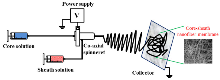
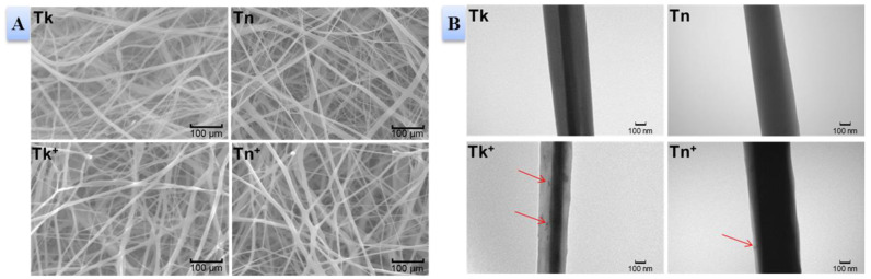
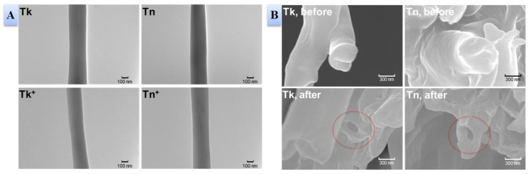
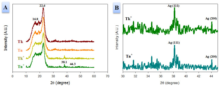
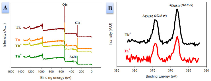
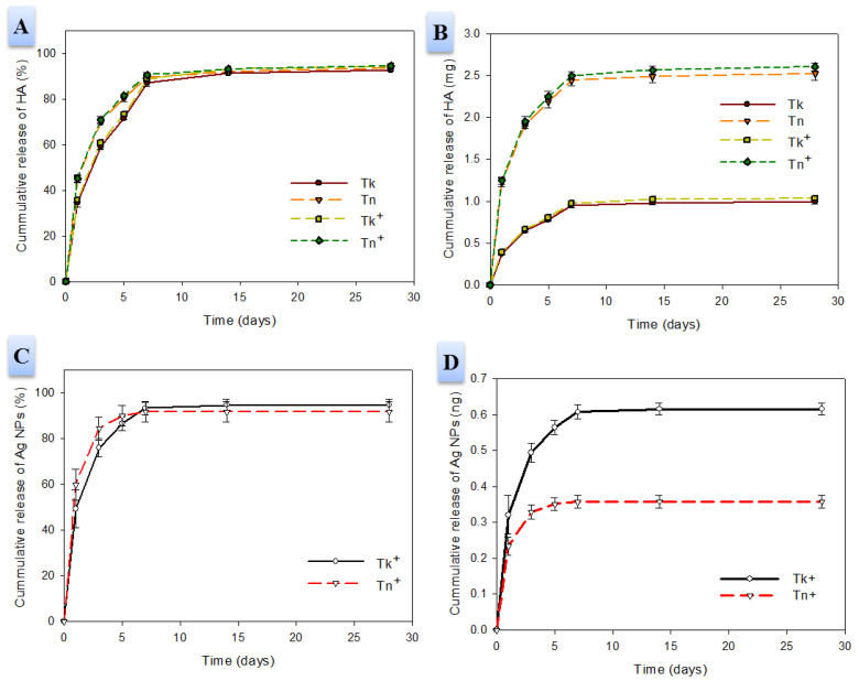
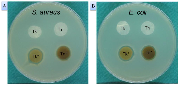
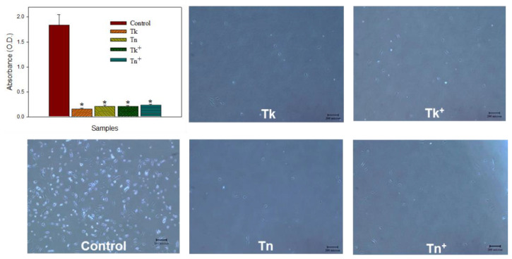
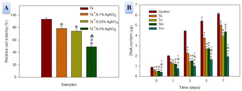
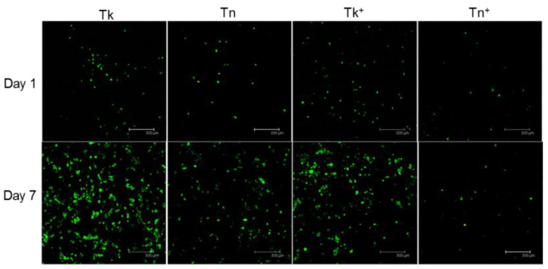
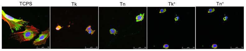
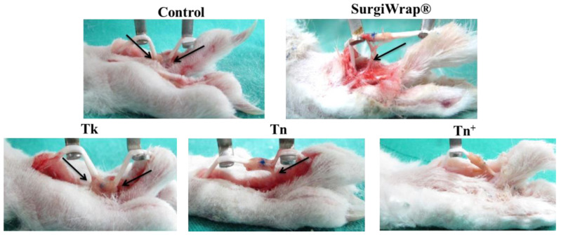
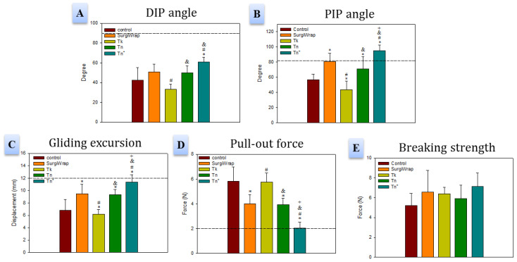
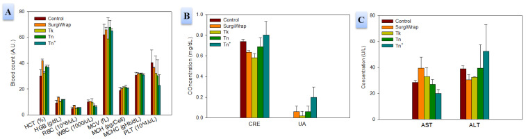
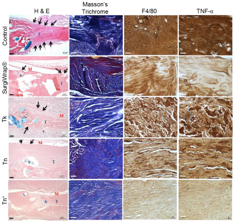
In this study, we prepared core-sheath nanofiber membranes (CSNFMs) with silver nanoparticles (Ag NPs) embedding in the polylactic acid (PLA) nanofiber sheath and hyaluronic acid (HA) in the nanofiber core. The PLA/Ag NPs sheath provides mechanical support as well as anti-bacterial and anti-inflammatory properties. The controlled release of HA from the core could exert anti-adhesion effects to promote tendon sliding while reducing fibroblast attachment. From the microfibrous structural nature of CSNFMs, they function as barrier membranes to reduce fibroblast penetration without hampering nutrient transports to prevent post-operative peritendinous adhesion. As the anti-adhesion efficacy will depend on release rate of HA from the core as well as Ag NP from the sheath, we fabricated CSNFMs of comparable fiber diameter, but with thick (Tk) or thin (Tn) sheath. Similar CSNFMs with thick (Tk+) and thin (Tn+) sheath but with embedded Ag NPs in the sheath were also prepared. The physico-chemical properties of the barrier membranes were characterized in details, together with their biological response including cell penetration, cell attachment and proliferation, and cytotoxicity. Peritendinous anti-adhesion models in rabbits were used to test the efficacy of CSNFMs as anti-adhesion barriers, from gross observation, histology, and biomechanical tests. Overall, the CSNFM with thin-sheath and Ag NPs (Tn+) shows antibacterial activity with low cytotoxicity, prevents fibroblast penetration, and exerts the highest efficacy in reducing fibroblast attachment in vitro. From in vivo studies, the Tn+ membrane also shows significant improvement in preventing peritendinous adhesions as well as anti-inflammatory efficacy, compared with Tk and Tn CSNFMs and a commercial adhesion barrier film (SurgiWrap®) made from PLA.

Keywords: anti-adhesion; core-sheath nanofibers; electrospinning; hyaluronic acid; polylactic acid; silver nanoparticles.


Conflict of interest statement

The authors declare no conflict of interest. The funders had no role in the design of the study; in the collection, analyses, or interpretation of data; in the writing of the manuscript, or in the decision to publish the results.

Figures

Scheme 1
Scheme 1
The schematic diagram for preparation of core-sheath nanofiber membranes (CSNFMs). The sheath spinning solution contains polylactic acid in methylene chloride/N,N’-dimethylformamide. For silver nanoparticles (Ag NPs)-embedded CSNFMs, the sheath spinning solution contains AgNO3, which is exposed to UV light beforehand to form Ag NPs. The core spinning solution contains hyaluronic acid and polyethylene oxide in formic acid.
Figure 1
Figure 1
The scanning electron microscopy (SEM) (A, bar = 100 μm) and transmission electron microcopy (TEM) (B, bar = 100 nm) images of Tk, Tn, Tk+, and Tn+ core-sheath nanofiber membranes (CSNFMs). The red arrows in (B) indicate Ag NPs in the sheath.
Figure 2
Figure 2
(A) The transmission electron microcopy (TEM) images of Tk, Tn, Tk+, and Tn+ CSNFMs after immersion in PBS for two weeks (bar = 100 nm). (B) The field-emission scanning electron microscopy (FE-SEM) images of Tk and Tn core-sheath nanofiber membranes (CSNFMs) before and after immersion in PBS for two weeks (bar = 300 nm). The red circles indicate the hollow core structure after eluting HA from the core.
Figure 3
Figure 3
The X-ray diffraction (XRD) analysis of Tk, Tn, Tk+, and Tn+ CSNFMs (A) and high resolution XRD spectra of Tk+ and Tn+ CSNFMs (B).
Figure 4
Figure 4
The X-ray photoelectron spectroscopy (XPS) analysis of Tk, Tn, Tk+, and Tn+ CSNFMs (A) and high resolution XPS spectra of Tk+, and Tn+ CSNFMs (B).
Figure 5
Figure 5
The cumulative release percentage (A) and cumulative released mass (B) of HA. The cumulative release percentage (C) and cumulative released mass (D) of Ag NPs.
Figure 6
Figure 6
The antibacterial activity of CSNFMs determined from the bacteriostatic rings for S. aureus (A) and E. coli (B). A disk shaped membrane with 1.5 cm diameter is used in the study and the weight of Ag NPs in Tn+ and TK+ are ~0.0195 and ~0.0325 mg, respectively.
Figure 7
Figure 7
The penetration of 3T3 fibroblasts through a CSNFM in 24 h by determining penetrated cell number from solution absorbance with MTS assays, and direct microscopic observation of penetrated cells (bar = 200 μm). The control is without a CSNFM. * p < 0.05 compared with control.
Figure 8
Figure 8
(A) The cytotoxicity tests of CSNFMs using the indirect contact method. * p < 0.05 compared with Tk; # p < 0.05 compared with Tk+/0.1% AgNO3; & p < 0.05 compared with Tk+/0.25% AgNO3. (B) The attachment and proliferation of 3T3 fibroblasts on control (TCPS) and CSNFMs from DNA assays. * p < 0.05 compared with control; # p < 0.05 compared with Tk; & p < 0.05 compared with Tn; + p < 0.05 compared with Tk+.
Figure 9
Figure 9
The Live/Dead staining of fibroblasts on CSNFMs by calcein AM and propidium iodide for live (green) and dead (red) cells after cultured for one and seven days and observed by confocal laser scanning microscopy. Bar = 300 μm.
Figure 10
Figure 10
The cytoskeletal F-actin (red) distribution and vinculin focal adhesion protein (green) expression of fibroblasts after cultured on control (TCPS) and different CSNFMs for one day and examined under a confocal laser scanning microscope. The cell nuclei were counterstained with DAPI (blue). Bar = 50 μm.
Figure 11
Figure 11
The rabbit flexor tendons observed three weeks post-operatively. Note the substantial peritendinous adhesions (black arrows) in the control and Tk groups, with fewer adhesions observed in the SurgiWrap® and Tn groups. Peritendinous adhesions are absent in the Tn+ group.
Figure 12
Figure 12
The assessment of peritendinous adhesions three weeks post-operation for distal interphalangeal (DIP) flexion angle (A), proximal interphalangeal (PIP) flexion angle (B), tendon gliding distance (C), and pull-out force (D). The dotted line represents the average value found for normal flexor digitorum profundus tendons. (E) Comparison of the breaking strength of healed tendons at three weeks. * p < 0.05 compared with control; # p < 0.05 compared with SurgiWrap®; & p < 0.05 compared with Tk; + p < 0.05 compared with Tn. The data are expressed as mean ± standard deviation (n = 8).
Figure 13
Figure 13
The blood testing results at one week postoperatively from complete blood count (A), renal (B), and liver (C) functions assessments. HCT, hematocrit; HGB, hemoglobin; RBC, red blood cell; WBC, white blood cell; MCV, mean corpuscular volume; MCH, mean corpuscular hemoglobin; MCHC: mean corpuscular hemoglobin concentration; PLT, platelet; CRE, creatinine; UA, uric acid; AST, aspartate aminotransferase; ALT, alanine aminotransferase. The data are expressed as mean ± standard deviation (n = 8).
Figure 14
Figure 14
The histological analysis of untreated tendons (control group), tendons treated with SurgiWrap®, and tendons treated with Tk, Tn, or Tn+ CSNFMs. The hematoxylin-eosin (H&E) staining shows adhesion tissues around repaired tendon (black arrows) as well the position of suture (S), tendon (T), and membrane (M) (bar = 200 μm). The Masson’s trichrome stain and immunohistochemical (IHC) staining of TNF-α and F4/80 are included (bar = 50 μm).

References

    1. Beyene R.T., Kavalukas S.L., Barbul A. Intra-abdominal adhesions: Anatomy, physiology, pathophysiology, and treatment. Curr. Probl. Surg. 2015;52:271–319. doi: 10.1067/j.cpsurg.2015.05.001. - DOI - PubMed
    1. Dy C.J., Hernandez-Soria A., Ma Y., Roberts T.R., Daluiski A. Complications After Flexor Tendon Repair: A Systematic Review and Meta-Analysis. J. Hand Surg. 2012;37:543–551. doi: 10.1016/j.jhsa.2011.11.006. - DOI - PubMed
    1. Titan A.L., Foster D.S., Chang J., Longaker M.T. Flexor Tendon: Development, Healing, Adhesion Formation, and Contributing Growth Factors. Plast. Reconstr. Surg. 2019;144:639e–647e. doi: 10.1097/PRS.0000000000006048. - DOI - PMC - PubMed
    1. Evanko S.P., Potter-Perigo S., Petty L.J., Workman G.A., Wight T.N. Hyaluronan Controls the Deposition of Fibronectin and Collagen and Modulates TGF-β1 Induction of Lung Myofibroblasts. Matrix Biol. 2015;42:74–92. doi: 10.1016/j.matbio.2014.12.001. - DOI - PMC - PubMed
    1. de Melo F., Carrijo A., Hong K., Trumbic B., Vercesi F., Waldorf H.A., Zenker S. Minimally Invasive Aesthetic Treatment of the Face and Neck Using Combinations of a PCL-Based Collagen Stimulator, PLLA/PLGA Suspension Sutures, and Cross-Linked Hyaluronic Acid. Clin. Cosmet. Investig. Dermatol. 2020;13:333–344. doi: 10.2147/CCID.S248280. - DOI - PMC - PubMed

MeSH terms

LinkOut - more resources