. 2021 Aug 17;22(16):8834.
doi: 10.3390/ijms22168834.

Sex-Specific Effects of Plastic Caging in Murine Viral Myocarditis

Affiliations

Sex-Specific Effects of Plastic Caging in Murine Viral Myocarditis

Katelyn A Bruno et al. Int J Mol Sci. .

Abstract

Background: Myocarditis is an inflammatory heart disease caused by viral infections that can lead to heart failure, and occurs more often in men than women. Since animal studies have shown that myocarditis is influenced by sex hormones, we hypothesized that endocrine disruptors, which interfere with natural hormones, may play a role in the progression of the disease. The human population is exposed to the endocrine disruptor bisphenol A (BPA) from plastics, such as water bottles and plastic food containers.

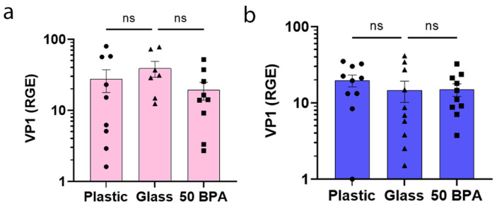
Methods: Male and female adult BALB/c mice were housed in plastic versus glass caging, or exposed to BPA in drinking water versus control water. Myocarditis was induced with coxsackievirus B3 on day 0, and the endpoints were assessed on day 10 post infection.

Results: We found that male BALB/c mice that were exposed to plastic caging had increased myocarditis due to complement activation and elevated numbers of macrophages and neutrophils, whereas females had elevated mast cell activation and fibrosis.

Conclusions: These findings show that housing mice in traditional plastic caging increases viral myocarditis in males and females, but using sex-specific immune mechanisms.

Keywords: bisphenol A; coxsackievirus B3; endocrine disruptors; myocarditis; sex differences.


Conflict of interest statement

The authors declare no conflict of interest.

Figures

Figure 1
Figure 1
Plastic caging increases myocardial inflammation during acute myocarditis in male BALB/c mice. (a) Female (pink) and (b) male (blue) BALB/c mice were given normal drinking water (drinking water that did not contain BPA) for 2 weeks and housed either in glass cages/water bottles (glass) or plastic cages/water bottles (plastic) with soy-free food and bedding, or BALB/c mice housed in glass cages/water bottles with soy-free food and bedding were either given normal drinking water (drinking water that did not contain BPA) (glass) or 250 μg BPA/L water (50 BPA) for 2 weeks. After BPA exposure, mice received an intraperitoneal (i.p.) injection with 103 plaque-forming units (PFU) of CVB3 on day 0 and harvested at day 10 post infection (pi). Myocarditis was assessed as % inflammation compared to the total size of the heart section with H&E using a microscope grid. Data shown as scatter plot and mean +/−SEM using one-way ANOVA with Holm–Šídák’s multiple comparisons test with 9–10 mice/group (* p < 0.05). (c) Representative H&E images of cardiac inflammation in plastic caging (plastic), glass caging (glass) or with 50 BPA in drinking water (50 BPA) (magnification 200×, scale bar 100 μm).
Figure 2
Figure 2
Plastic caging or BPA did not increase cardiac VP1 viral gene expression during acute myocarditis. (a) Female (pink) and (b) male (blue) adult BALB/c mice were given normal drinking water (drinking water that did not contain BPA) for 2 weeks and housed in glass cages/water bottles (glass) or plastic cages/water bottles (plastic) caging with soy-free food and bedding. BALB/c mice housed in glass cages/water bottles with soy-free food and bedding received either normal drinking water (drinking water that did not contain BPA) (glass) or 250 μg BPA/L water (50 BPA) for 2 weeks. Mice were injected i.p. with 103 PFU of CVB3 on day 0 and harvested at day 10 pi. Relative gene expression (RGE) of CVB3 VP1 levels in the heart were determined using qRT-PCR compared to the housekeeping gene hypoxanthine phosphoribosyltransferase (Hprt). Data shown as scatter plot and mean +/−SEM using one-way ANOVA with Holm–Šídák’s multiple comparisons test with 7–10 mice/group.
Figure 3
Figure 3
Plastic caging increases macrophage and neutrophil markers during acute myocarditis in BALB/c males. Male BALB/c mice were given normal drinking water (drinking water that did not contain BPA) for 2 weeks and housed either in glass cages/water bottles (glass) or plastic cages/water bottles (plastic) with soy-free food and bedding. BALB/c mice were housed in glass cages/water bottles with soy-free food and bedding and administered either normal drinking water (drinking water that did not contain BPA) (glass) or 250 μg BPA/L water (50 BPA) for 2 weeks. Mice were injected i.p. with 103 PFU of CVB3 on day 0 and harvested at day 10 pi. Relative gene expression (RGE) of whole hearts by qRT-PCR was used to assess (a) CD45 (total lymphocytes), (b) CD11b+ cells (i.e., macrophages, neutrophils, mast cells), (c) F4/80+ macrophages, (d) GR1 (neutrophils), (e) CD14 (part of the TLR4 signaling complex), compared to the housekeeping gene Hprt. Data shown as scatter plot and mean +/−SEM using one-way ANOVA with Holm–Šídák’s multiple comparisons test with 9–10 mice/group (* p < 0.05), (** p < 0.01).
Figure 4
Figure 4
Plastic caging has no effect on expression of T-cell markers during acute myocarditis in BALB/c males. Male BALB/c mice were given normal drinking water (drinking water that did not contain BPA) for 2 weeks and housed either in glass cages/water bottles (glass) or plastic cages/water bottles (plastic) with soy-free food and bedding. BALB/c mice housed in glass cages/water bottles with soy-free food and bedding were given either normal drinking water (drinking water that did not contain BPA) (glass) or 250 μg BPA/L (50 BPA) in drinking water for 2 weeks. Mice were injected i.p. with 103 PFU of CVB3 on day 0 and harvested at day 10 pi. Relative gene expression (RGE) was used in whole hearts by qRT-PCR to assess (a) CD3+ T cells, (b) CD4+ T cells and (c) CD8+ T cells compared to the housekeeping gene Hprt. Data shown as scatter plot and mean +/−SEM using one-way ANOVA with Holm–Šídák’s multiple comparisons test with 10 mice/group (* p < 0.05), (** p < 0.01).
Figure 5
Figure 5
Plastic caging increases macrophages and neutrophils during acute myocarditis in BALB/c males. Male BALB/c mice were given normal drinking water (drinking water that did not contain BPA) for 2 weeks and housed either in glass cages/water bottles (glass) or plastic cages/water bottles (plastic) with soy-free food and bedding. BALB/c mice were housed in glass cages/water bottles with soy-free food and bedding and administered either normal drinking water (drinking water that did not contain BPA) (glass) or 250 μg BPA/L water (50 BPA) for 2 weeks. Mice were injected i.p. with 103 PFU of CVB3 on day 0 and harvested at day 10 pi. Five micron sections were stained with antibodies against (a) CD45 (total lymphocytes), (b) CD11b+ cells (i.e., macrophages, neutrophils, mast cells), (c) F4/80+ macrophages, (d) GR1 (neutrophils), (e) CD3+ T cells, and positive pixels were compared to total positive and negative pixels to determine % positive. Data shown as scatter plot and mean +/−SEM using one-way ANOVA with Holm–Šídák’s multiple comparisons test for 17–19 mice/group (* p < 0.05), (** p < 0.01), (**** p < 0.0001). Representative images of positive-stained cells (brown) in plastic caging (plastic), glass caging (glass) and 50 BPA in drinking water (50 BPA) in glass cages shown for (a) CD45, (b) CD11b, (c) F4/80, (d) GR1, and (e) CD3. Red arrow points to brown GR1-positive cells (magnification 200×, scale bar 200 μm).
Figure 6
Figure 6
Plastic caging increases the mast cell marker cKit during acute myocarditis in BALB/c females, but decreases its expression in males. (a) Female (pink) and (b) male (blue) BALB/c mice were given normal drinking water (drinking water that did not contain BPA) for 2 weeks and housed either in glass cages/water bottles (glass) or plastic cages/water bottles (plastic) with soy-free food and bedding. BALB/c mice housed in glass cages/water bottles with soy-free food and bedding were given either normal drinking water (drinking water that did not contain BPA) (glass) or 250 μg BPA/L water (50 BPA) for 2 weeks. Mice were injected i.p. with 103 PFU CVB3 on day 0 and harvested at day 10 pi. The cKit levels in the heart were determined by qRT-PCR for relative gene expression (RGE) compared to the housekeeping gene Hprt. Data shown as scatter plot and mean +/−SEM using one-way ANOVA with Holm–Šídák’s multiple comparisons test with 10 mice/group (* p < 0.05), (**** p < 0.0001).
Figure 7
Figure 7
Plastic caging increases pericardial mast cell numbers and degranulation in females during myocarditis. Female BALB/c mice (pink) were given (ae) normal drinking water (drinking water that did not contain BPA) for 2 weeks and housed either in glass cages/water bottles (glass) or plastic cages/water bottles (plastic) with soy-free food and bedding. (fj) BALB/c mice housed in glass cages/water bottles with soy-free food and bedding were given either normal drinking water (drinking water that did not contain BPA) (glass) or 250 μg BPA/L water (50 BPA) for 2 weeks. Mice were injected i.p. with 103 PFU of CVB3 on day 0 and harvested at day 10 pi. Mast cell scoring was completed using the 40× high-power objective on a compound microscope. Mast cells were counted and categorized according to whether they were degranulated or not and by their location as pericardial, myocardial or near vessels. Data shown as scatter plot and mean +/−SEM using a Student’s t-test with 9–10 mice/group (* p < 0.05), (** p < 0.01), (*** p < 0.001), (**** p < 0.0001). Representative histology images of mast cells (purple, arrow). Representative images in plastic caging (plastic), glass caging (glass), or 50 BPA in drinking water (50 BPA) in glass cages show mast cells associated with the (k) pericardium, (l) myocardium or (m) vessels (magnification 400×, scale bar 60 μm).
Figure 8
Figure 8
Plastic caging does not alter mast cell numbers or degranulation in males during myocarditis. Male BALB/c mice (blue) were given normal drinking water (drinking water that did not contain BPA) for 2 weeks and housed either in glass cages/water bottles (glass) or plastic cages/water bottles (plastic) with soy-free food and bedding. BALB/c mice housed in glass cages/water bottles with soy-free food and bedding were given either normal drinking water (drinking water that did not contain BPA) (0 BPA/glass) or 250 μg BPA/L water (50 BPA) for 2 weeks. Mice were injected i.p. with 103 PFU of CVB3 on day 0 and harvested at day 10 pi. Mast cell scoring was completed using the 40× high-power objective on a compound microscope. (ae) Mast cells were counted and categorized according to whether they were degranulated (small blue granules can be observed released from the cell at high power) or not and by their location as pericardial, myocardial or near vessels. Data shown as scatter plot and mean +/−SEM using one-way ANOVA with Holm–Šídák’s multiple comparisons test with 10 mice/group (** p < 0.01), (**** p < 0.0001). (f,g) Representative histology images of mast cells (purple, arrow). Representative images in plastic caging (plastic), glass caging (glass) or with 50 BPA in drinking water in glass cages (50 BPA) show (f) mast cells that are not degranulating (NOT) versus (g) degranulating mast cells (degran) with identifiable mast cell granules outside of the cell (magnification 400×, scale bar 60 μm).
Figure 9
Figure 9
Plastic caging associated with pericardial mast cell degranulation in females, but not in males, where mast cells degranulate regardless of plastic exposure. (a,c) Female (pink) and (b,d) male (blue) BALB/c mice were given (a,b) normal drinking water (drinking water that did not contain BPA) for 2 weeks and housed either in glass cages/water bottles (glass) or plastic cages/water bottles (plastic) with soy-free food and bedding. (c,d) BALB/c mice housed in glass cages/water bottles with soy-free food and bedding were given either normal drinking water (drinking water that did not contain BPA) (glass) or 250 μg BPA/L water (50 BPA) in glass cages for 2 weeks. Mice were injected i.p. with 103 PFU of CVB3 on day 0 and harvested at day 10 pi. Mast cell scoring was completed using the 40× high-power objective on a compound microscope. Mast cells were counted and categorized according to whether they were degranulated or not and by their location as pericardial. Data shown as scatter plot and mean +/−SEM using a Student’s t-test with 9–10 mice/group (* p < 0.05), (** p < 0.01), (*** p < 0.001).
Figure 10
Figure 10
Plastic caging increases collagen gene expression and fibrosis in the heart of females, but not male mice during myocarditis. (a,b) Female (pink) and (c,d) male (blue) BALB/c mice were given normal drinking water (drinking water that did not contain BPA) for 2 weeks and housed either in glass cages/water bottles (glass) or plastic cages/water bottles (plastic) with soy-free food and bedding. BALB/c mice housed in glass caging with soy-free food and bedding were given either normal drinking water (drinking water that did not contain BPA) (0 BPA/glass) for 2 weeks or 250 μg BPA/L water (50 BPA). Mice were injected i.p. with 103 PFU of CVB3 i.p. on day 0 and harvested at day 10 pi. (a,c) Collagen 1 levels in the heart were measured using qRT-PCR to determine relative gene expression (RGE) of collagen I compared to the housekeeping gene Hprt. (b,d) Fibrosis was assessed as the % fibrosis compared to the total size of the heart section using Sirius Red-stained sections and a microscope eyepiece grid. Data shown as scatter plot and mean +/−SEM using one-way ANOVA with Holm–Šídák’s multiple comparisons test with 8–12 mice/group (* p < 0.05), (** p < 0.01), (*** p < 0.001). Representative images of fibrosis detected with Sirius Red (arrow) shown in the (e) pericardium, (f) myocardium or (g) vessels (magnification 400×, scale bar 60 μm).
Figure 11
Figure 11
Plastic caging decreases ERα and AR and increases ERβ expression in the heart of males, but has no effect on ERs in females during myocarditis. (ad) Female (pink) and (eh) male (blue) BALB/c mice were given normal drinking water (drinking water that did not contain BPA) for 2 weeks and housed either in glass cages/water bottles (glass) or plastic cages/water bottles (plastic) with soy-free food and bedding. BALB/c mice housed in glass cages/water bottles with soy-free food and bedding were given either normal drinking water (drinking water that did not contain BPA) (0 BPA/glass) or 250 μg BPA/L water (50 BPA) for 2 weeks. Mice were injected i.p. with 103 PFU of CVB3 on day 0 and harvested at day 10 pi. Relative gene expression (RGE) was assessed in whole hearts by qRT-PCR. (* p < 0.05), (** p < 0.01). Expression of (a,e) ERα, (b,f) ERβ, (c,g) ERRγ, and (d,h) AR were assessed compared to the housekeeping gene Hprt. Data shown as scatter plot and mean +/−SEM using one-way ANOVA with Holm–Šídák’s multiple comparisons test with 8–10 mice/group.
Figure 12
Figure 12
Plastic caging increases complement markers in males with myocarditis, but not in females. Male (blue) BALB/c mice were given normal drinking water (drinking water that did not contain BPA) for 2 weeks and housed either in glass cages/water bottles (glass) or plastic cages/water bottles (plastic) with soy-free food and bedding. BALB/c mice housed in glass cages/water bottles with soy-free food and bedding were given either normal drinking water (drinking water that did not contain BPA) (0 BPA/glass) or 250 μg BPA/L water (50 BPA) for 2 weeks. Mice were injected i.p. with 103 PFU of CVB3 on day 0 and harvested at day 10 pi. Relative gene expression (RGE) was used in whole hearts by qRT-PCR to assess (a) complement receptor 3 (CR3), (b) complement component 3 antagonist receptor 1 (C3aR1), (c) complement component C4b, or (d) complement component 5 antagonist receptor 1 (C5aR1), compared to the housekeeping gene Hprt. Data shown as scatter plot and mean +/−SEM using one-way ANOVA with Holm–Šídák’s multiple comparisons test with 10 mice/group (* p < 0.05), (** p < 0.01).

References

    1. Pauschinger M., Phan M.D., Doerner A., Kuehl U., Schwimmbeck P.L., Poller W., Kandolf R., Schultheiss H.P. Enteroviral RNA replication in the myocardium of patients with left ventricular dysfunction and clinically suspected myocarditis. Circulation. 1999;99:889–895. doi: 10.1161/01.CIR.99.7.889. - DOI - PubMed
    1. Kuhl U., Pauschinger M., Seeberg B., Lassner D., Noutsias M., Poller W., Schultheiss H.P. Viral persistence in the myocardium is associated with progressive cardiac dysfunction. Circulation. 2005;112:1965–1970. doi: 10.1161/CIRCULATIONAHA.105.548156. - DOI - PubMed
    1. Fairweather D., Stafford K.A., Sung Y.K. Update on coxsackievirus B3 myocarditis. Curr. Opin. Rheumatol. 2012;24:401–407. doi: 10.1097/BOR.0b013e328353372d. - DOI - PMC - PubMed
    1. Sawalha K., Abozenah M., Kadado A.J., Battisha A., Al-Akchar M., Salerno C., Hernandez-Montfort J., Islam A.M. Systematic review of COVID-19 related myocarditis: Insights on management and outcome. Cardiovasc Revasc. Med. 2021;23:107–113. doi: 10.1016/j.carrev.2020.08.028. - DOI - PMC - PubMed
    1. Long B., Brady W.J., Koyfman A., Gottlieb M. Cardiovascular complications in COVID-19. Am. J. Emerg. Med. 2020;38:1504–1507. doi: 10.1016/j.ajem.2020.04.048. - DOI - PMC - PubMed

LinkOut - more resources