Review
. 2021 Aug 19;22(16):8942.
doi: 10.3390/ijms22168942.

Radiotherapy of High-Grade Gliomas: First Half of 2021 Update with Special Reference to Radiosensitization Studies

Affiliations
Review

Radiotherapy of High-Grade Gliomas: First Half of 2021 Update with Special Reference to Radiosensitization Studies

Guido Frosina. Int J Mol Sci. .

Abstract

Albeit the effort to develop targeted therapies for patients with high-grade gliomas (WHO grades III and IV) is evidenced by hundreds of current clinical trials, radiation remains one of the few effective therapeutic options for them. This review article analyzes the updates on the topic "radiotherapy of high-grade gliomas" during the period 1 January 2021-30 June 2021. The high number of articles retrieved in PubMed using the search terms ("gliom* and radio*") and manually selected for relevance indicates the feverish research currently ongoing on the subject. During the last semester, significant advances were provided in both the preclinical and clinical settings concerning the diagnosis and prognosis of high-grade gliomas, their radioresistance, and the inevitable side effects of their treatment with radiation. The novel information concerning tumor radiosensitization was of special interest in terms of therapeutic perspective and was discussed in detail.

Keywords: high-grade-glioma; radiotherapy; sensitization; update.


Conflict of interest statement

The author declares no conflict of interest.

Figures
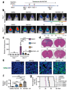
Figure 1
Figure 1
Inhibiting DNA-PKcs in combination with IR improved GB treatment. (ac) In vivo bioluminescent imaging of orthotopic GB xenografts derived from H2S GICs expressing luciferase in immunocompromised NSG mice. Seven days after GIC transplantation, NSG mice were treated with DNA-PKcs inhibitor (NU7441) or DMSO (control) by intraperitoneal injection and/or irradiated (IR; 2 × 2 Gy) on day 9 after GIC transplantation. Representative images (b) of the four indicated treatment groups on day 7 (before treatment) and day 17 (after treatment) and quantifications (c) of tumor growth in mouse brains are shown. Data are means ± SD. n = 5 mice per group. ANOVA analysis was used to assess the significance. **, p < 0.01, ***, p < 0.001, and ****, p < 0.0001. (d) Representative images showing H&E staining of cross-sections of mouse brains bearing GIC-derived tumors from four groups of mice treated with NU7441, IR (irradiation), NU7441 + IR, or DMSO (control). Scale bar, 2 mm. (e,f) TUNEL assay detecting apoptosis (green) in the GB xenografts treated with the DNA-PKcs inhibitor NU7441, irradiation (IR), NU7441 + IR, or DMSO (control) and counterstained with DAPI (blue) to indicate nuclei (e). Quantifications of cell apoptosis from four treatment groups (f). Scale bars, 50 μm. ANOVA analysis was used to assess the significance. Data are means ± SD. n = 4 independent experiments (about 200 cells per arm). ****, p < 0.0001. (g) Kaplan–Meier survival curves of mice intracranially transplanted with H2S GSCs and treated with the DNA-PKcs inhibitor NU7441, irradiation (IR), NU7441 + IR, or DMSO (control). n = 5 animals per group. Log-rank analysis was used. Control versus NU7441, p = 0.0017; control versus IR, p = 0.0017; control versus NU7441 + IR, p = 0.0017; NU7441 versus NU7441 + IR, p = 0.0018; and IR versus NU7441 + IR, p = 0.0025 (modified after [67] with permission).
Figure 2
Figure 2
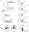
Hematopoietic stem cell (HSC) gene therapy targeting transforming growth factor beta (TGFβ) in combination with IR results in a long-term protection against intracranial GB. (a) Survival of mice bearing GL261 intracranial tumors. The table below the graph shows group sizes at the beginning (day 0) and number of animals at risk (e.g., animals that are alive) for indicated days. Pooled data from two independent experiments are shown. Statistical significance was determined by a two-tailed log-rank test. (b) Growth curves for individual tumors as obtained by bioluminescence imaging, displaying total flux in photons per second (p/s). (c) Mice that rejected GL261 tumors following the first intracranial implantation of cancer cells plus therapy were rechallenged by a second intracranial implantation of GL261 cells at 90 days post-tumor rejection (red and green lines). Mice that have received transplantation of MMP14: GFP-transduced HSCs (gray lines) or naïve mice (black lines) were implanted with GL261 cells intracranially for the first time and used as controls. Tumor growth was quantified by bioluminescence imaging as in (b). (d,e) Representative dot plots showing analysis of CD8+ and CD4+ T cells (d) and quantification of immune cells (e) in intracranial tumors (control mice as specified in c) or brain area corresponding to the cancer cell implantation site (mice that have rejected tumors for the second time in the combination therapy group) by flow cytometry at 3 weeks post-cancer cell implantation. Statistical significance was determined by two-tailed t-test (n = 3 and 4, respectively, in first and second experiment for control group; n = 2 per experiment for rechallenged long-term survivors from the combination therapy group) (modified after [68] with permission).
Figure 3
Figure 3
Tumor genotype dictates radiosensitization after Atm deletion in primary brainstem glioma models. (a). Deletion of Atm in p53-deficient gliomas improves tumor response to IR. Kaplan–Meier plot of overall survival in tumor-bearing mice after three daily fractions of 10 Gy RT delivered to the whole brain. *, p < 0.05 by log rank test. (b). Deletion of Atm does not radiosensitize p53 wild-type gliomas. Kaplan–Meier plot of overall survival in tumor-bearing mice after three daily fractions of 10 Gy RT delivered to the whole brain. *, p < 0.05 by t test (2 tailed). (c,d). p53 wild-type gliomas with intact ATM function are sensitive to IR. Kaplan–Meier plot of overall survival in (c) p53-deficient tumor-bearing nPAFL/+ mice or (d) p53 wild-type tumor-bearing nIAFL/+ mice that received no RT or were treated with three daily fractions of 10 Gy to the whole brain. (e). Kaplan–Meier plot comparing the survival of mice with p53 wild-type and p53-deficient gliomas treated with fractionated RT. The animals included in these survival curves are the same animals from the survival studies in panels (a) and (b). *, p < 0.05 by log rank test. (f). p53 signaling mediates radiosensitivity of Ink4a/Arf-deficient gliomas. Kaplan–Meier plot comparing the survival of nPAFL/+, nIAFL/+, and nPIAFL/+ mice after treatment with three daily fractions of 10 Gy delivered to the whole brain. Control nPAFL/+ and nIAFL/+ curves were taken from panels (a) and (b). *, p < 0.05 by log rank test. (gi). Deletion of Atm improves the radiation response of gliomas driven by loss of p53 and Ink4a/Arf. (g) Kaplan–Meier plot of overall survival in tumor-bearing mice after tumor detection. (h) Kaplan–Meier plot of overall survival in tumor-bearing mice after three daily fractions of 10 Gy RT delivered to the whole brain. (i) Kaplan–Meier plot comparing the survival of mice with p53-deficient gliomas and gliomas lacking both p53 and Ink4a/Arf that were treated with fractionated RT. Control nPAFL/+ and nPAFL/FL curves were taken from panels (a,b). *, p < 0.05 by t test (two-tailed) when comparing the mean of two groups or log rank test when comparing survival curves. Tumor genotypes/phenotypes. nPAFL/+, nestinTVA p53FL/FL AtmFL/+ lacking p53; nPAFL/FL, nestinTVA p53FL/FL AtmFL/FL lacking both p53 and ATM; nIAFL/+, nestinTVA Ink4a/ArfFL/FL AtmFL/+/lacking Ink4a/Arf; nIAFL/FL, nestinTVA Ink4a/ArfFL/FL AtmFL/FL/lacking both Ink4a/Arf. and Atm; nPIAFL/+, nestinTVA p53FL/FL Ink4a/ArfFL/FL AtmFL/+ lacking both p53 and Ink4a/Arf (modified after [69] with permission).
Figure 4
Figure 4
p53 modulates RT fraction size sensitivity in normal and malignant cells. (ad). Split-dose recovery is not observed in primary fibroblast with loss of functional p53. (ad). Transformed Li-Fraumeni fibroblasts MDAH041 were exposed to either acute or split-dose IR with indicated doses. (a) Colony survival assay confirms loss of split-dose recovery (white triangle, single acute dose, and gray triangle represents split dose IR). (b) Schema for (c) Western blot analysis showing the expression levels of total p53, p21, and loading control GAPDH and (d) colony survival of p53 siRNA knockdown in 1BR hTERT cells for the indicated period. UT is untreated, mock represents cells treated with DharmaFECT1 transfection reagent, and Scr is the ON-TARGETplus non-targeting control scramble. RF, the ratio of split-dose to single dose survival, has been compared for each experimental condition. (e,f). Split-dose recovery is lost in tumor cell lines with mutant p53. Colony survival of tumor cell lines A2780 WT (e,f) A2780 E6 after exposure to either acute or daily fractionated IR with indicated doses. Top panel in each histogram shows the experimental schema, white triangle represents single acute dose and gray triangle 1 Gy daily fractions. Post IR (6 h) cells were trypsinized and pooled with cells collected from media, plated, and allowed to form colonies. A significant increase in split-dose recovery is observed in p53 WT A2780 (e) but not in mut p53 A2780 E6 (f) (modified after [72] with permission).

References

    1. McCutcheon I.E., Preul M.C. Historical Perspective on Surgery and Survival with Glioblastoma: How Far have we Come? World Neurosurg. 2021;149:148–168. doi: 10.1016/j.wneu.2021.02.047. - DOI - PubMed
    1. Omuro A., DeAngelis L.M. Glioblastoma and Other Malignant Gliomas: A Clinical Review. JAMA. 2013;310:1842–1850. doi: 10.1001/jama.2013.280319. - DOI - PubMed
    1. Khan I., Mahfooz S., Elbasan E.B., Karacam B., Oztanir M.N., Hatiboglu M.A. Targeting Glioblastoma: The Current State of Different Therapeutic Approaches. Curr. Neuropharmacol. 2021;19:1. doi: 10.2174/1570159X19666210113152108. - DOI - PMC - PubMed
    1. Stupp R., Mason W.P., van den Bent M.J., Weller M., Fisher B., Taphoorn M.J., Belanger K., Brandes A.A., Marosi C., Bogdahn U., et al. Radiotherapy Plus Concomitant and Adjuvant Temozolomide for Glioblastoma. N. Engl. J. Med. 2005;352:987–996. doi: 10.1056/NEJMoa043330. - DOI - PubMed
    1. Oronsky B., Reid T.R., Oronsky A., Sandhu N., Knox S.J. A Review of Newly Diagnosed Glioblastoma. Front. Oncol. 2021;10:574012. doi: 10.3389/fonc.2020.574012. - DOI - PMC - PubMed

Substances