. 2021 Aug 27;19(1):167.
doi: 10.1186/s12915-021-01077-2.

Unexpected organellar locations of ESCRT machinery in Giardia intestinalis and complex evolutionary dynamics spanning the transition to parasitism in the lineage Fornicata

Affiliations

Unexpected organellar locations of ESCRT machinery in Giardia intestinalis and complex evolutionary dynamics spanning the transition to parasitism in the lineage Fornicata

Shweta V Pipaliya et al. BMC Biol. .

Abstract

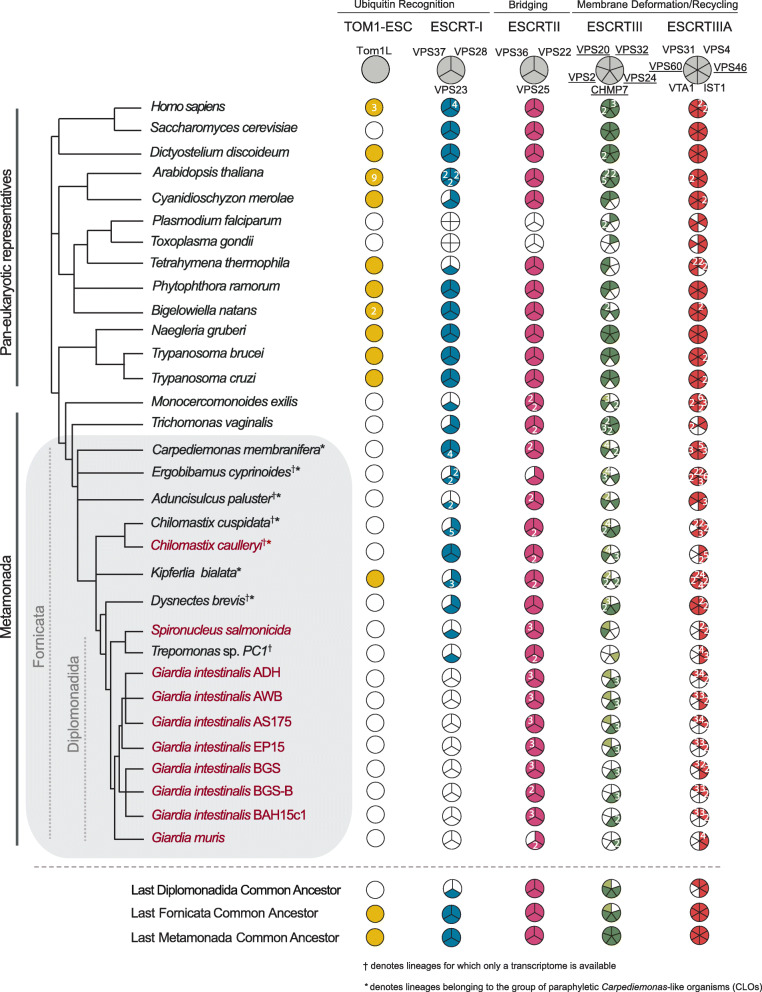
Background: Comparing a parasitic lineage to its free-living relatives is a powerful way to understand how that evolutionary transition to parasitism occurred. Giardia intestinalis (Fornicata) is a leading cause of gastrointestinal disease world-wide and is famous for its unusual complement of cellular compartments, such as having peripheral vacuoles instead of typical endosomal compartments. Endocytosis plays an important role in Giardia's pathogenesis. Endosomal sorting complexes required for transport (ESCRT) are membrane-deforming proteins associated with the late endosome/multivesicular body (MVB). MVBs are ill-defined in G. intestinalis, and roles for identified ESCRT-related proteins are not fully understood in the context of its unique endocytic system. Furthermore, components thought to be required for full ESCRT functionality have not yet been documented in this species.

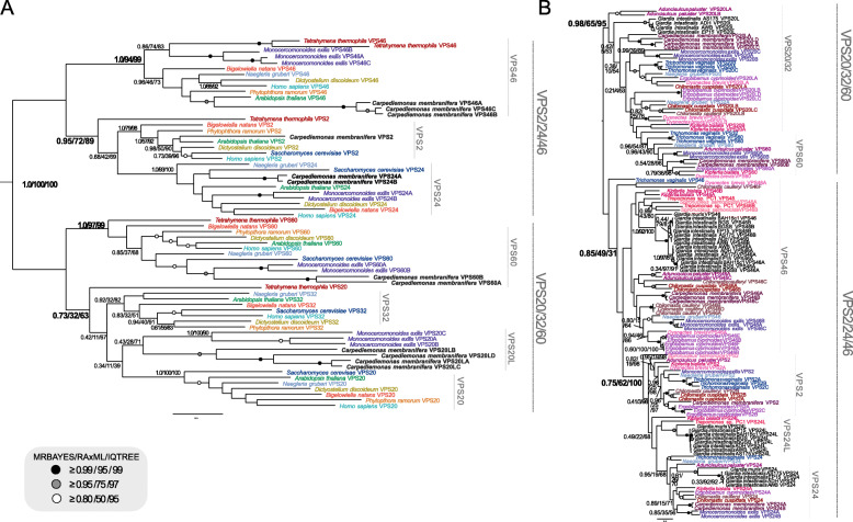
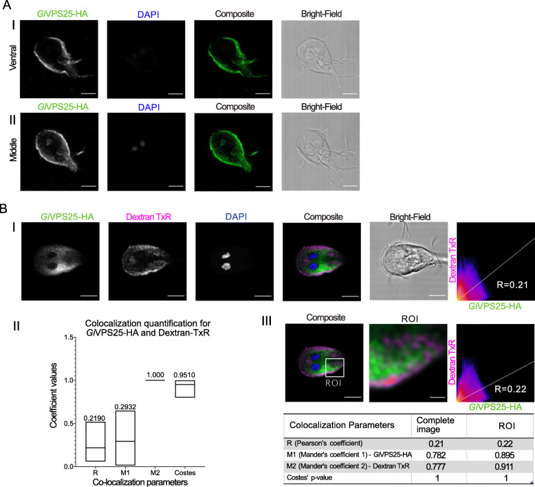
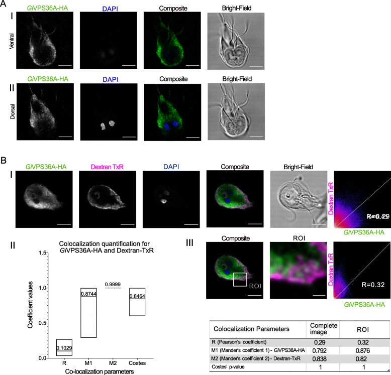
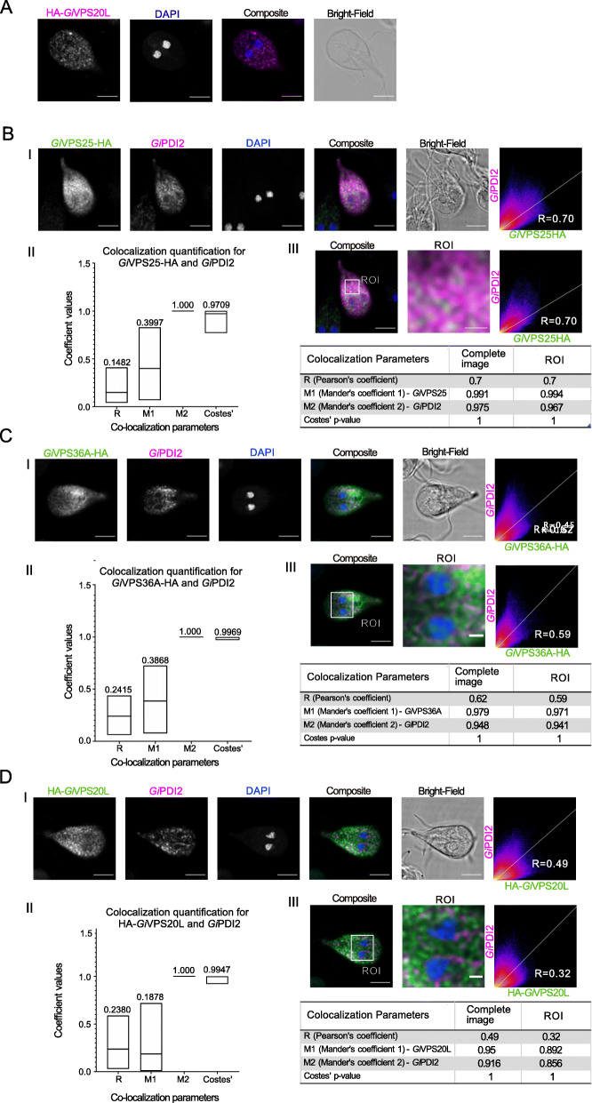
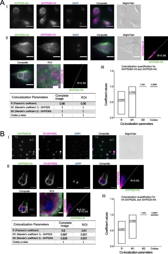
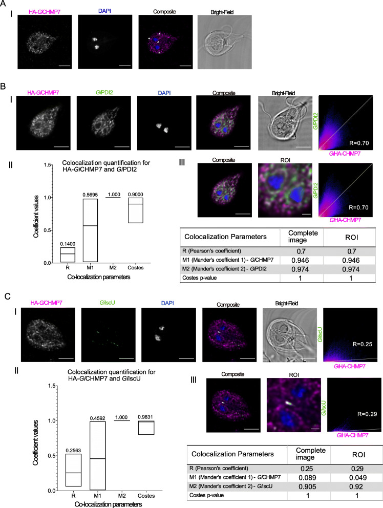
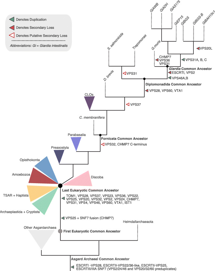
Results: We used genomic and transcriptomic data from several Fornicata species to clarify the evolutionary genome streamlining observed in Giardia, as well as to detect any divergent orthologs of the Fornicata ESCRT subunits. We observed differences in the ESCRT machinery complement between Giardia strains. Microscopy-based investigations of key components of ESCRT machinery such as GiVPS36 and GiVPS25 link them to peripheral vacuoles, highlighting these organelles as simplified MVB equivalents. Unexpectedly, we show ESCRT components associated with the endoplasmic reticulum and, for the first time, mitosomes. Finally, we identified the rare ESCRT component CHMP7 in several fornicate representatives, including Giardia and show that contrary to current understanding, CHMP7 evolved from a gene fusion of VPS25 and SNF7 domains, prior to the last eukaryotic common ancestor, over 1.5 billion years ago.

Conclusions: Our findings show that ESCRT machinery in G. intestinalis is far more varied and complete than previously thought, associates to multiple cellular locations, and presents changes in ESCRT complement which pre-date adoption of a parasitic lifestyle.

Keywords: ESCRT; Endomembrane; Evolutionary Cell Biology; Excavata; Giardia; PV; Parasitism.


Conflict of interest statement

The authors declare that they have no competing interests.

Figures

Fig. 1
Fig. 1
Distribution of ESCRT components within Fornicata. Coulson plot summary depicting ESCRT complement identified in Fornicata genomes and transcriptomes in comparison to pan-eukaryotic representatives. Filled sectors indicate subunits with solidified orthology determined using both comparative genomics and phylogenetics. Numbers within individual sectors represent multiple paralogues. Light coloured sectors indicate ambiguous phylogenetic classification but confirmed reciprocal blast orthology. Taxa for which genomes were available and examined are indicated in plain text whereas lineages where only transcriptomes are indicated with a superscript symbol. Lineages belonging to the paraphyletic group of Carpediemonas-like organisms indicated with an asterisks. Parasitic fornicates are indicated in burgundy. Paralagous ESCRTIII and ESCRTIIIA subunits possessing the SNF7 domain with common evolutionary origins, and for downstream phylogenetic investigations, are underlined. Of important note, only inferences regarding gene presence, not absences, can be made conclusively in the lineages for which only a transcriptome is available
Fig. 2
Fig. 2
Phylogenetic analysis of ESCRTs in Fornicata. A Phylogenetic analyses of the ESCRTIII/IIIA SNF7 families in Fornicata. Identified ESCRTIII/IIIA SNF7 components from the basal Carpediemonas membranifera as a landmark representative for Fornicata were subject to phylogenetic classification. Two of the identified SNF7 sequences from Carpediemonas membranifera clustered clearly with VPS60 whereas the remainder neither strongly grouped with VPS20 or VPS32 and therefore were determined to be VPS20L proteins in all tree topologies. Carpediemonas membranifera was also determined to have VPS2, VPS24, and VP46 with strong backbone clade support for two paralogs of VPS24 (1.0/100/100) and three paralogs of VPS46 (1.0/100/100). B A Fornicata-specific tree with well characterized Discoba and metamonad representatives. Monocercomonoides exilis, Trichomonas vaginalis, and Naegleria gruberi as well as newly characterized sequences from Carpediemonas membranifera were used to classify SNF7 components in all CLOs and diplomonads. Similar to Carpediemonas, no clear grouping of SNF7 sequences from CLOs within the VPS20 or VPS32 clade was observed and therefore was also classified as VPS20L. Only sequences from Giardia AWB, ADH, and EP15 formed a group within this clade and therefore were also determined to be VPS20L. VPS2 family proteins identified in the diplomonads grouped with both VPS24 and VPS46 with duplication event pointing in Giardia sp. VPS46 yielding two paralogues, VPS46A and VPS46B. An additional set of SNF7 family proteins from Giardia AWB, ADH, and EP15 grouped with excavate and CLO VPS24 proteins therefore were determined to be VPS24-like proteins. However, an additional set of SNF7 proteins from all Giardia lineages formed a separate sister clade and therefore also termed to be VPS24. Trees were rooted between the VPS20/32/60 and VPS2/24/46 as previously determined by [21]
Fig. 3
Fig. 3
Characterization of GiVPS25-HA subcellular location. A Trophozoite cell periphery and cytosol. (I) Ventral and (II) middle optical slice of transgenic Giardia trophozoite expressing epitope-tagged GiVPS25-HA (green). All images were obtained using Laser Scanning Confocal Microscopy. All scale bars: 5 μm. B PVs. (I) Immunofluorescence assay of transgenic Giardia trophozoite labelled for epitope-tagged GiVPS25-HA (green) and Dextran-TexasRed (magenta). (II) Distribution of co-localization parameters for GiVPS25-HA and Dextran-TexasRed labeling from ≥ 15 analysed cells. Mean values for each parameter are indicated. (III) Signal overlap analysis and co-localization coefficients calculated for all slices of the sample either for the whole cell or ROI. Scale bars: composite 5 μm and ROI 1 μm. All images were obtained using Laser Scanning Confocal Microscopy
Fig. 4
Fig. 4
Characterization of GiVPS36A-HA subcellular location. A Cell periphery and cytosol. (I) Ventral and (II) dorsal optical slices of transgenic Giardia trophozoite expressing GiVPS36A-HA. All images were obtained using Laser Scanning Confocal Microscopy. All scale bars: 5 μm. B PVs. (I) Immunofluorescence assay of transgenic Giardia trophozoites labelled for GiVPS36-HA (green) after incubation with Dextran-TxR (magenta). (II) Distribution of co-localization parameters for GiVPS36-HA and Dextran-TexasRed labeling from ≥ 15 analysed cells. Mean values for each parameter are indicated. (III) Signal overlap analysis and co-localization coefficients calculated for all slices of the sample either for the whole cell or ROI. Scale bars: composite 5 μm and ROI 1 μm. All images were obtained using Laser Scanning Confocal Microscopy
Fig. 5
Fig. 5
Co-labelling of GiVPS25-HA, GiVPS36A-HA, and GiHA-VPS20L with ER membrane marker GiPDI2. A HA-GiVPS20L is found in the cytosol and punctate structures. Scale bars: 5 μm. B–D Panels I: Co-labelling of PDI2 (magenta) in cells expressing either B GiVPS25-HA (green), C GiVPS36A-HA (green), or D HA-GiVPS20L (green). B–D Panels II: Mean values from ≥ 15 analysed cells for each parameter are indicated. B–D Panels III: Signal overlap analysis and co-localization coefficients calculated for all slices of the sample either for the whole cell or ROI. Scale bars: composite 5 μm and ROI 1 μm. All images were obtained using Laser Scanning Confocal Microscopy
Fig. 6
Fig. 6
Co-expression of epitope-tagged GiVPS25 with either GiVPS20L or GiVPS36. Microscopy analysis of cells co-expressing GiVPS25HA (green) with either A GiVPS36A-V5 (magenta) or B GiV5-VPS20L (magenta). Panels I: representative cell images and percentage of co-labeling. Panels II: Signal overlap analysis in both whole cells and regions of interest (ROI). Scale bars: (I) 10 μm, (II whole cell) 5 μm, and (II-ROI) 1 μm. Panels III: Mean values from ≥ 15 analysed cells for each parameter are indicated. All images were obtained using Laser Scanning Confocal Microscopy
Fig. 7
Fig. 7
Ab initio homology-based structural analysis of the CHMP7 N-terminus. A Homology-based protein structural analysis of the CHMP7 N-terminus from various pan-eukaryotic representatives carried out using iTASSER ab initio structural prediction program where considerable structural similarity between the ESCRTII-VPS25 and CHMP7 N-termini. B Proposed evolution as determined by homology searching, structural analyses, and phylogenetic analysis (Supplementary Table S2, Supplementary Figures S8) of the pan-eukaryotic CHMP7 protein prior to the last eukaryotic common ancestor which consisted of an evolutionary fusion event between a pre-LECA ESCRTII-VPS25 and ESCRTIII/IIIA-SNF7 progenitor protein
Fig. 8
Fig. 8
Characterization of GiCHMP7 subcellular location. A Immunofluorescence assays of HA-GiCHMP7-expressing cells yield a diffused punctate pattern with elements of perinuclear ER staining (arrowhead). Scale bars: 5 μm. B (I) Co-labelling of HA-GiCHMP7 (magenta) -expressing cells with GiPDI2 (green). (II) Distribution of co-localization parameters for HA-GiCHMP7 and GiPDI2 labeling from ≥ 15 analysed cells. Mean values for each parameter are indicated. (III) Signal overlap analysis for all slices of the sample either for the whole cell or ROI. C HA-GiCHMP7 is associated to Giardia mitosomes. (I) Co-labelling of HA-GiCHMP7 (magenta) -expressing cells with GiIscU (green). (II) Distribution of co-localization parameters for HA-GiCHMP7 and GiIscU labeling from ≥ 15 analysed cells. Mean values for each parameter are indicated. (III) Signal overlap analysis for all slices of the sample either for the whole cell or ROI. Scale bar: composite 5 μm and ROI 1 μm. All images were obtained using Laser Scanning Confocal Microscopy
Fig. 9
Fig. 9
Proposed ESCRT evolution in Fornicata. Progenitor ESCRT complexes are present in Asgard archaea and duplications into the specific subunits is inferred to have occurred between the First Eukaryotic Common Ancestor and the Last Eukaryotic Common Ancestor which possessed a full complement of the ESCRT subunits. Proposed ESCRT losses in Fornicata inferred previously only using Giardia intestinalis are transient with some losses potentially pre-dating the Last Fornicata Common Ancestor [21, 25]. The most prominent of this being loss in CHMP7 c-terminus SNF7 domain and a canonical VPS32. Examination of diplomonad lineages, specifically genomic data, increases our confidence in additional losses also occurring with progression into parasitism most notable within the ESCRTI machinery with complete loss occurring in the Giardia common ancestor likely associated with a loss in the canonical MVB morphology. Speculative losses indicated as unfilled dotted arrows whereas instances of likely true gene absence depicted as solid filled arrows

Similar articles

Cited by

References

    1. Lanata CF, Fischer-Walker CL, Olascoaga AC, Torres CX, Aryee MJ, Black RE, et al. Global causes of diarrheal disease mortality in children < 5 years of age: a systematic review. PLoS ONE. 2013;8(9):e72788. doi: 10.1371/journal.pone.0072788. - DOI - PMC - PubMed
    1. Faso C, Hehl AB. Membrane trafficking and organelle biogenesis in Giardia lamblia: Use it or lose it. Int J Parasitol. 2011;41(5):471–480. doi: 10.1016/j.ijpara.2010.12.014. - DOI - PubMed
    1. Allain T, Buret AG. Chapter Five - Pathogenesis and post-infectious complications in giardiasis. In: Ortega-Pierres MG, editor. Advances in Parasitology. Academic Press; 2020. pp. 173–199. - PubMed
    1. Faso C, Hehl AB. Advances in parasitology. Elsevier; 2019. A cytonaut’s guide to protein trafficking in Giardia lamblia; pp. 105–127. - PubMed
    1. Gargantini PR, Serradell M Del C, Ríos DN, Tenaglia AH, Luján HD. Antigenic variation in the intestinal parasite Giardia lamblia. Host-Microbe Interact Parasitesfungiviruses. 2016;32:52–58. - PubMed

Publication types

MeSH terms

Substances