

Application of the "telescopic rod" in a combined surgical technique for the treatment of congenital pseudarthrosis of the tibia in children
- PMID: 34446041
- PMCID: PMC8390273
- DOI: 10.1186/s13018-021-02649-2
Application of the "telescopic rod" in a combined surgical technique for the treatment of congenital pseudarthrosis of the tibia in children
Abstract
Background: The current surgical treatment of choice is the combination surgical technique, involving tibial intramedullary fixation to maintain the mechanical axis and mechanical stability of tibial pseudarthrosis. In traditional combined surgery, the Williams rod is often used. Long-term intramedullary fixation of the foot and ankle will affect the ankle joint function of children. The intramedullary rod is relatively shorter due to the growth of the distal tibia. In addition, there are some complications such as epiphyseal bone bridge and high-arched foot. The use of a telescopic intramedullary rod may avoid these complications.
Purposes: To investigate the initial effect of the "telescopic rod" in a combined surgical technique for the treatment of congenital pseudarthrosis of the tibia in children.
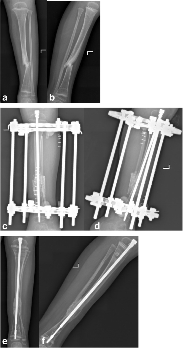
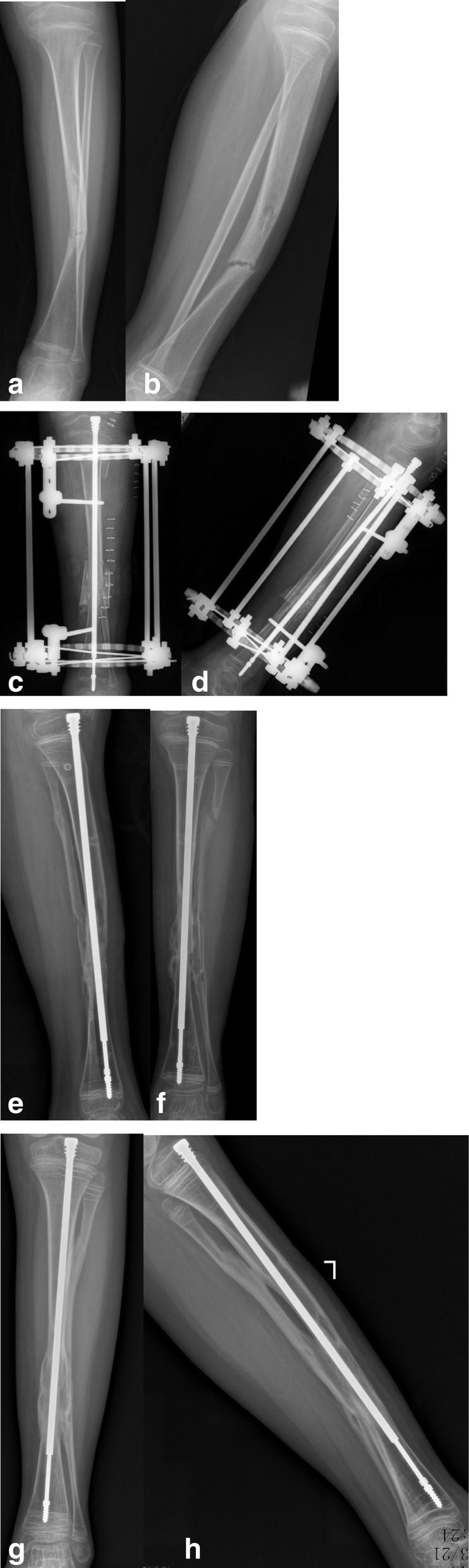
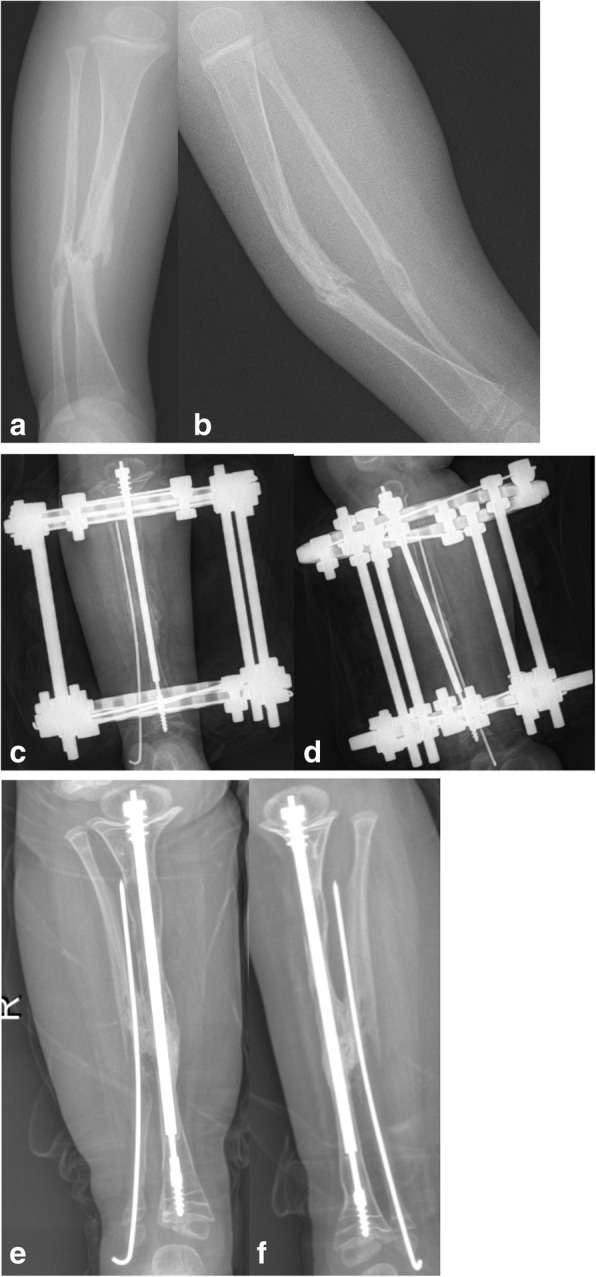
Methods: A retrospective study including 15 patients with Crawford type IV CPT who were treated using a combined surgical technique and the telescopic rod from January 2017 to May 2018. The average age at the time of surgery was 43.3 months (16-126 months). Of the 15 patients, 7 had proximal tibia dysplasia and 12 exhibited neurofibromatosis type 1. The combined surgical technique using the telescopic rod included the excision of pseudarthrosis, intramedullary rod insertion, installation of Ilizarov's fixator, tibia-fibular cross union, and wrapping autogenic iliac bone graft. The incidence of refracture, ankle valgus, tibial valgus, and limb length discrepancy (LLD) in patients were investigated.
Results: All patients achieved primary union with an average follow-up time of 37.3 months (26-42 months). The mean primary union time was 4.5 months (4.0-5.6 months). Nine cases showed LLD (60%), with an average limb length of 1.1 cm (0.5-2.0 cm). Ankle valgus, proximal tibial valgus, telescopic rod displacement, and epiphyseal plate tethering occurred in 1 case (6.6%) (18°), 3 cases (20%) (10°, 5°, and 6°, respectively), 6 cases (40%), and 2 cases (13%), respectively. There were no refractures during the follow-up periods.
Conclusion: Although there are complications such as intramedullary rod displacement while using the telescopic rod in a combined surgery, the primary healing rate of congenital pseudarthrosis of the tibia in children is high.
Keywords: Children; Congenital pseudarthrosis of tibia; Initial effect; Telescopic rod.
© 2021. The Author(s).
Conflict of interest statement
The authors declare that they have no competing interests.
Figures
References
-
- Liu YX, Mei HB, Zhu GH, He RG, Liu K, Tang J, Wu JY, Ye WH, Hu X, Tan Q, Yan A, Huang SX, Tan XQ, Lei T. Relationship between postoperative complications and fibular integrity in congenital pseudarthrosis of the tibia in children. World J Pediatr. 2017;13(3):261–266. doi: 10.1007/s12519-016-0074-2. - DOI - PubMed
-
- Liu YX, Mei HB, Liu K, et al. A new radiographic classification scheme of congenital pseudarthrosis of the tibia. Chin J Pediatr Surg. 2016;37(1):29–33. doi: 10.3760/cma.j.issn.0253-3006.2016.01.007. - DOI
MeSH terms
Supplementary concepts
LinkOut - more resources
Full Text Sources
Research Materials
