. 2021 Aug 31;118(35):e2106673118.
doi: 10.1073/pnas.2106673118.

SPATA33 localizes calcineurin to the mitochondria and regulates sperm motility in mice

Affiliations

SPATA33 localizes calcineurin to the mitochondria and regulates sperm motility in mice

Haruhiko Miyata et al. Proc Natl Acad Sci U S A. .

Abstract

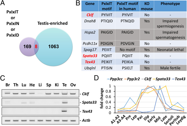
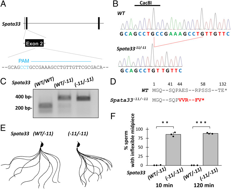
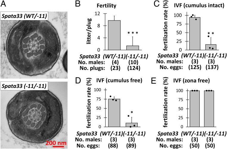
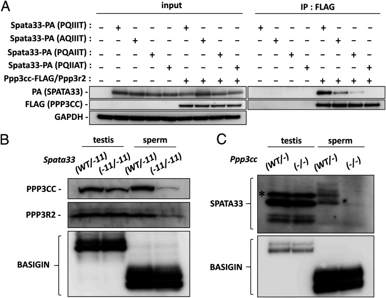
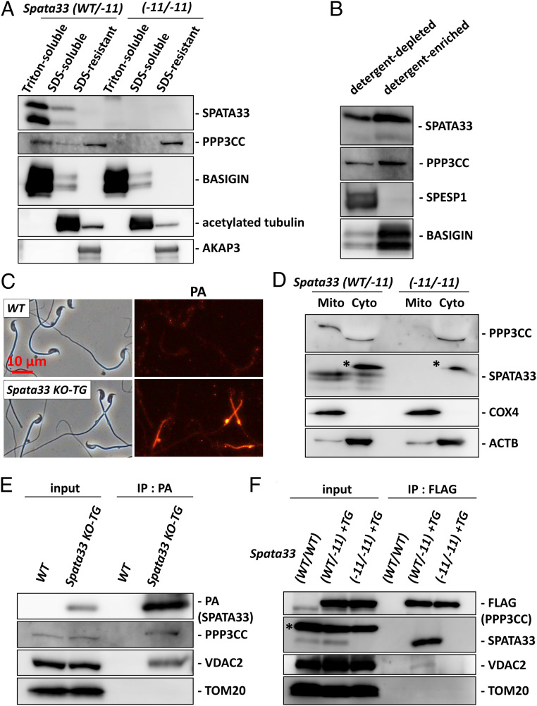
Calcineurin is a calcium-dependent phosphatase that plays roles in a variety of biological processes including immune responses. In spermatozoa, there is a testis-enriched calcineurin composed of PPP3CC and PPP3R2 (sperm calcineurin) that is essential for sperm motility and male fertility. Because sperm calcineurin has been proposed as a target for reversible male contraceptives, identifying proteins that interact with sperm calcineurin widens the choice for developing specific inhibitors. Here, by screening the calcineurin-interacting PxIxIT consensus motif in silico and analyzing the function of candidate proteins through the generation of gene-modified mice, we discovered that SPATA33 interacts with sperm calcineurin via a PQIIIT sequence. Spata33 knockout mice exhibit reduced sperm motility because of an inflexible midpiece, leading to impaired male fertility, which phenocopies Ppp3cc and Ppp3r2 knockout mice. Further analysis reveals that sperm calcineurin disappears from the mitochondria in the Spata33 knockout testis. In addition, immunoprecipitation analysis indicates that sperm calcineurin interacts with not only SPATA33 but also the mitochondrial protein VDAC2. These results indicate that SPATA33 localizes calcineurin to the mitochondria and regulates sperm motility.

Keywords: calcineurin; male fertility; mitochondria; sperm motility.


Conflict of interest statement

The authors declare no competing interest.

Figures

Fig. 1.
Fig. 1.
Screening proteins that interact with sperm calcineurin. (A) Mouse proteins with PxIxIT, PxIxIN, or PxIxID that are expressed strongly in the testis were searched. There are eight genes that are included in both categories. (B) Testis-enriched genes that possess PxIxIT motif. Cklf, Spata33, and Tex43 were analyzed further. (C) RT-PCR of Cklf, Spata33, and Tex43 using RNAs obtained from mouse various tissues. Actb was the control. Br: brain, Th: thymus, Lu: lung, He: heart, Li: liver, Sp: spleen, Ki: kidney, Te: testis, Ov: ovary. (D) In silico data analysis of Ppp3cc, Ppp3r2, Cklf, Spata33, and Tex43 expression level. A1-A2: A1 and A2 differentiating spermatogonia, A3-A4: A3, A4, In, and B differentiating spermatogonia, Pre: preleptotene spermatocytes, Lep: leptotene/zygotene spermatocytes, Pac: pachytene spermatocytes, Dip: diplotene/secondary spermatocytes, Early: early round spermatids, Mid: mid round spermatids, Late: late round spermatids, Sertoli: Sertoli cells, Peritub: peritubular myoid cells, and Leydig: Leydig cells.
Fig. 2.
Fig. 2.
SPATA33 is essential for midpiece flexibility. (A) CRISPR/Cas9 targeting scheme. gRNA was designed within exon 2. Protospacer adjacent motif is shown in blue. (B) Wave pattern sequence of Spata33. In mutants, 11-bp nucleotides were deleted, which disrupts a Cac8I restriction site. (C) Genotyping Spata33−11/−11 mice by Cac8I digestion. (D) The 11-bp deletion caused a A44V mutation with a premature stop codon introduced 14 amino acids later. (E) Flagellar waveforms were analyzed 10 min after incubation. Single frames throughout one beating cycle were superimposed. Spermatozoa from Spata33−11/−11 mice exhibit inflexible midpieces. (F) The percentage of the spermatozoa with an inflexible midpiece at 10 and 120 min after sperm suspension. n = 3 males each for Spata33WT/−11 and Spata33−11/−11 mice. **P < 0.01 and ***P < 0.001 (Student's t test).
Fig. 3.
Fig. 3.
In vivo and in vitro fertility of Spata33−11/−11 mice. (A) No obvious ultrastructural abnormalities were observed in the midpiece of Spata33−11/−11 spermatozoa. (B) Number of litters born per plug detected. (C) In vitro fertilization (IVF) with cumulus-intact oocytes. (D) IVF with cumulus-free oocytes. (E) IVF with zona-free oocytes. The impaired fertility of Spata33−11/−11 mice was rescued. *P < 0.05, **P  <  0.01, and ***P  <  0.001 (Student's t test).
Fig. 4.
Fig. 4.
SPATA33 interacts with sperm calcineurin via PQIIIT sequence. (A) Spata33-PA (PQIIIT) or mutated Spata33-PA (AQIIIT, PQAIIT, or PQIIAT) was coexpressed with Ppp3cc-FLAG/Ppp3r2 in HEK293T cells, and immunoprecipitation (IP) with FLAG M2 antibody was performed. GAPDH was the control. (B) PPP3CC and PPP3R2 decreased in the spermatozoa from Spata33−11/−11 mice. BASIGIN was the control. (C) SPATA33 decreased in the spermatozoa from Ppp3cc−/− mice. BASIGIN was the control. The asterisk indicates a nonspecific band.
Fig. 5.
Fig. 5.
SPATA33 localizes sperm calcineurin to the mitochondria. (A) Fractionation of mouse spermatozoa. SPATA33 was found in the Triton-soluble fraction. PPP3CC is found in both the Triton-soluble and SDS-resistant fraction. Only PPP3CC in the Triton-soluble fraction was depleted in Spata33−11/−11 mice. BASIGIN, acetylated tubulin, and AKAP3 were used as makers for the Triton-soluble, SDS-soluble, and SDS-resistant fractions, respectively. (B) Phase separation of Triton X-114 extracts of spermatozoa. SPATA33 and PPP3CC were enriched in the detergent-enriched phase. SPESP1 is a marker for the detergent-depleted fraction, whereas BASIGIN is a marker for the detergent-enriched fraction. (C) SPATA33-PA localization was analyzed using Spata33-PA TG mice. SPATA33-PA is localized in the midpiece. (D) Separation of mitochondrial and cytosolic fractions of testicular proteins. Both PPP3CC and SPATA33 were found in the mitochondrial fraction. PPP3CC was depleted in the mitochondrial fraction of Spata33−11/−11 mice. PPP3CC bands in the cytosolic fraction were shifted due to the existence of a large amount of protein (probably tubulin). The asterisks indicate nonspecific bands. (E) Immunoprecipitation with anti-PA antibody was performed using testis lysates of SPATA33-PA TG mice. PPP3CC and VDAC2 were immunoprecipitated with SPATA33-PA. (F) Immunoprecipitation with anti-FLAG antibody was performed using testis lysates of Ppp3cc-FLAG TG mice. SPATA33 and VDAC2 were immunoprecipitated with PPP3CC-FLAG. The asterisk indicates a nonspecific band.

References

    1. Eddy E. M., Toshimori K., O’Brien D. A., Fibrous sheath of mammalian spermatozoa. Microsc. Res. Tech. 61, 103–115 (2003). - PubMed
    1. Miyata H., Morohoshi A., Ikawa M., Analysis of the sperm flagellar axoneme using gene-modified mice. Exp. Anim. 69, 374–381 (2020). - PMC - PubMed
    1. Klee C. B., Ren H., Wang X., Regulation of the calmodulin-stimulated protein phosphatase, calcineurin. J. Biol. Chem. 273, 13367–13370 (1998). - PubMed
    1. Rusnak F., Mertz P., Calcineurin: Form and function. Physiol. Rev. 80, 1483–1521 (2000). - PubMed
    1. Miyata H., et al. ., Sperm calcineurin inhibition prevents mouse fertility with implications for male contraceptive. Science 350, 442–445 (2015). - PubMed

Publication types

Substances