. 2021 Aug 26;11(1):17251.
doi: 10.1038/s41598-021-96225-7.

Optical coherence tomographic angiography study of perfusion recovery after surgical lowering of intraocular pressure

Affiliations

Optical coherence tomographic angiography study of perfusion recovery after surgical lowering of intraocular pressure

Liang Liu et al. Sci Rep. .

Abstract

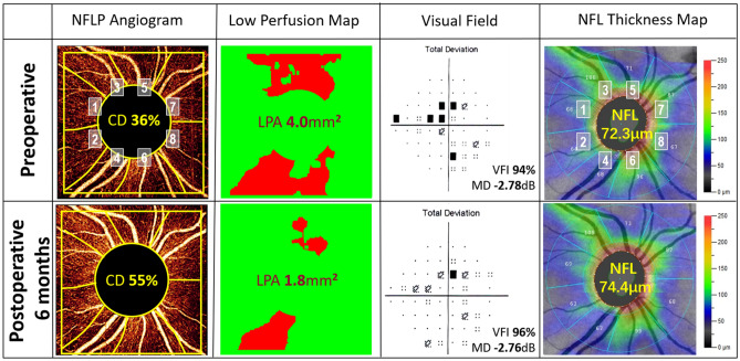
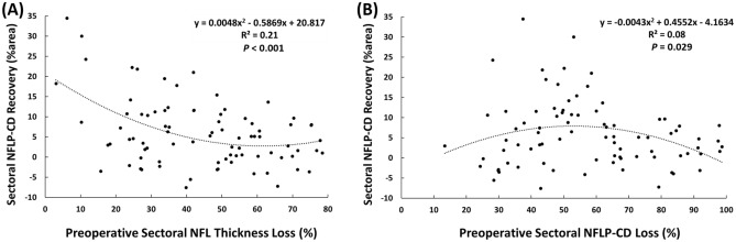
We investigated the time and location of retinal perfusion recovery after surgical intraocular pressure (IOP) lowering in glaucoma by using optical coherent tomography angiography (OCTA). Seventeen patients were analyzed. The 4.5 × 4.5-mm OCTA scans centered on the disc were performed preoperatively and postoperatively at 6 weeks, 3 months, and 6 months. The peripapillary retinal nerve fiber layer (NFL) thickness, NFL plexus capillary density (NFLP-CD) and visual field (VF) were measured overall and in 8 corresponding sectors. The low-perfusion area (LPA) was used to assess the cumulative area where local NFLP-CD was significantly below normal. At 6 months, the average IOP decreased 5.3 mmHg (P = 0.004), LPA decreased by 15% (P = 0.005), and NFLP-CD improved by 12% (P < 0.001). The NFL thickness and VF mean deviation didn't change significantly at any time point. Among the sectors with significant preoperative NFLP-CD loss, the recovery at 6 months was greatest in sectors with minimal preoperative NFL thinning (P < 0.001). In conclusion, surgical IOP lowering may improve NFLP capillary perfusion after 6 months. The perfusion recovery tended to occur in areas with minimal NFL thinning at baseline. OCTA parameters may have potential usefulness as pharmacodynamic biomarkers for glaucoma therapy.


Conflict of interest statement

OHSU, Yali Jia, Ou Tan and David Huang have financial interest in Optovue, Inc., a company that may have a commercial interest in the results of this research and technology. These potential conflicts of interest have been reviewed and are managed by OHSU. The other authors do not report any potential financial conflicts of interest.

Figures

Figure 1
Figure 1
A mildly glaucomatous eye showed retinal perfusion recovery at 6 months after trabeculectomy. The nerve fiber layer plexus (NFLP) angiogram was divided into 8 corresponding sectors according to an extended Garway-Heath scheme. This participant had lower IOP (from 16 to 13 mmHg) and less ocular antihypertensive medications use (from 3 to 0) at 6 months postoperatively. The NFLP capillary density (CD) increased by 53%. The low-perfusion map showed the perfusion defects mainly in the sector 3, 4, 5 and 6. The low-perfusion area (LPA) became 55% smaller after surgery. The perfusion recovery occurred primarily in the sector 3 and sector 6, where the NFL thickness were less thinning than the sector 5 and sector 4. The visual field (VF) total deviation map showed shallower defects. The rim area increased from 1.10 to 1.21 mm2 and the cup/disc area ratio decreased from 0.30 to 0.27. However, the VF mean deviation (MD) and nerve fiber layer (NFL) thickness showed minimal improvement.
Figure 2
Figure 2
The recovery of nerve fiber layer plexus capillary density (NFLP-CD) 6 months after surgery was correlated with preoperative parameters on a sector basis. Sectors with significant preoperative NFLP-CD loss were analyzed. (A) The plot against preoperative nerve fiber layer (NFL) thickness loss showed that the perfusion recovery was greatest in sectors with minimal preoperative NFL thinning (P < 0.001). (B) Plot against preoperative NFLP-CD loss showed greater recovery was possible in sectors with moderate preoperative perfusion loss (P = 0.029).

References

    1. Tham YC, et al. Global prevalence of glaucoma and projections of glaucoma burden through 2040: A systematic review and meta-analysis. Ophthalmology. 2014;121:2081–2090. doi: 10.1016/j.ophtha.2014.05.013. - DOI - PubMed
    1. Eye Diseases Prevalence Research Group Prevalence of open-angle glaucoma among adults in the united states. Arch. Ophthalmol. 2004;122:532–538. doi: 10.1001/archopht.122.4.532. - DOI - PMC - PubMed
    1. Musch DC, et al. Visual field improvement in the collaborative initial glaucoma treatment study. Am. J. Ophthalmol. 2014;158:96–104. doi: 10.1016/j.ajo.2014.04.003. - DOI - PMC - PubMed
    1. Caprioli J, et al. Trabeculectomy can improve long-term visual function in glaucoma. Ophthalmology. 2016;123:117–128. doi: 10.1016/j.ophtha.2015.09.027. - DOI - PubMed
    1. Baril C, et al. Rates of glaucomatous visual field change after trabeculectomy. Br. J. Ophthalmol. 2017;101:874–878. doi: 10.1136/bjophthalmol-2016-308948. - DOI - PubMed

Publication types