Observational Study
. 2021 Dec;101(12):1561-1570.
doi: 10.1038/s41374-021-00653-y. Epub 2021 Aug 26.

Establishing standardized immune phenotyping of metastatic melanoma by digital pathology

Collaborators, Affiliations
Observational Study

Establishing standardized immune phenotyping of metastatic melanoma by digital pathology

Bettina Sobottka et al. Lab Invest. 2021 Dec.

Erratum in

Abstract

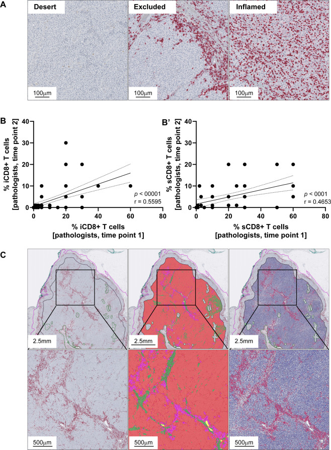
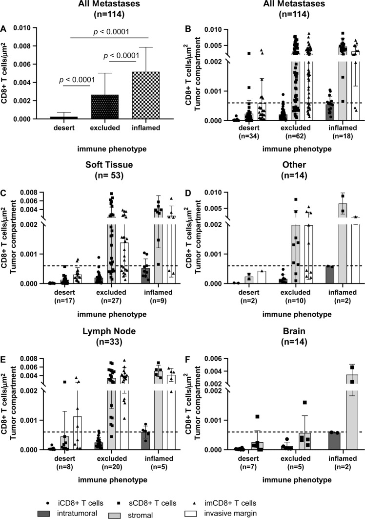
CD8+ tumor-infiltrating T cells can be regarded as one of the most relevant predictive biomarkers in immune-oncology. Highly infiltrated tumors, referred to as inflamed (clinically "hot"), show the most favorable response to immune checkpoint inhibitors in contrast to tumors with a scarce immune infiltrate called immune desert or excluded (clinically "cold"). Nevertheless, quantitative and reproducible methods examining their prevalence within tumors are lacking. We therefore established a computational diagnostic algorithm to quantitatively measure spatial densities of tumor-infiltrating CD8+ T cells by digital pathology within the three known tumor compartments as recommended by the International Immuno-Oncology Biomarker Working Group in 116 prospective metastatic melanomas of the Swiss Tumor Profiler cohort. Workflow robustness was confirmed in 33 samples of an independent retrospective validation cohort. The introduction of the intratumoral tumor center compartment proved to be most relevant for establishing an immune diagnosis in metastatic disease, independent of metastatic site. Cut-off values for reproducible classification were defined and successfully assigned densities into the respective immune diagnostic category in the validation cohort with high sensitivity, specificity, and precision. We provide a robust diagnostic algorithm based on intratumoral and stromal CD8+ T-cell densities in the tumor center compartment that translates spatial densities of tumor-infiltrating CD8+ T cells into the clinically relevant immune diagnostic categories "inflamed", "excluded", and "desert". The consideration of the intratumoral tumor center compartment allows immune phenotyping in the clinically highly relevant setting of metastatic lesions, even if the invasive margin compartment is not captured in biopsy material.


Conflict of interest statement

V.H.K. has served as an invited speaker on behalf of Indica Labs. H.M. is on advisory boards for Bayer, Astra Zeneca, Janssen, Roche, and Merck. R.D. reports intermittent, project-focused consulting and/or advisory relationships with Novartis, Merck Sharp & Dohme (MSD), Bristol Myers Squibb (BMS), Roche, Amgen, Takeda, Pierre Fabre, Sun Pharma, Sanofi, Catalym, Second Genome, Regeneron, and Alligator outside the submitted work. M.P.L. is a co-founder and shareholder of Oncobit AG and receives research funding from Novartis, Roche, and Molecular Partners. All other authors declare no conflict of interest.

Figures

Fig. 1
Fig. 1. Illustration of CD8+ T cells in relation to their immune phenotypes.
(A) Absence of T cells in immune desert tumors, accumulation of T cells at the invasive margin or in the intratumoral stroma without effective invasion in immune excluded tumors and infiltration of T cellsinto the tumor parenchyma in inflamed tumors. Correlation analysis of pathologists’ first and second semi-quantitative evaluation of the intratumoral CD8+ (iCD8+) T-cell (B) and stromal CD8+ (sCD8+) T-cell (B′) percentages; solid lines = best fit, dotted lines = error bars. Pathologist guided annotation (C) of the tumor border with 1 mm invasive margin (C, left) with exclusion of artefacts according to the recommendations by the working group. AI-based segmentation (C, middle) of melanoma metastases into tumor (red), inflamed stroma (purple), and desmoplastic stroma (green); exclusion of glass background, melanin pigment, hemorrhage, and necrosis. Cell segmentation andscoring at single cell resolution (C, right) evaluating CD8+ infiltration per μm2 Q9 in each compartment and tissue type (Color figure online).
Fig. 2
Fig. 2. Discovery cohort.
Densities of CD8+ T cells/μm2 in the discovery cohort independent of the tumor compartment (A). Total densities of CD8+ T cells differed significantly between immune desert, excluded, and inflamed tumors (A). Densities of CD8+ T cells/μm2 depicted according to their spatial distribution among the tumor compartments intratumoral (iCD8+ T cells; left; dark gray columns), stroma (sCD8+ T cells; middle; light gray columns), and invasive margin (imCD8+ T cells; right; white columns) in relation to the immune diagnosis and site of metastasis (BF); line at 0.06 CD8+ T cells/μm2.
Fig. 3
Fig. 3. Diagnostic algorithm.
Proposed diagnostic algorithm using a highly standardized and reproducible approach to define an immune diagnosis based on densities of tumor-infiltrating CD8+ T cells/μm2/ tumor compartment.
Fig. 4
Fig. 4. Validation cohort.
Linear regression and correlation analysis between the digitally assessed and classified immune diagnoses and the pathologists’ diagnoses in the validation cohort (A). While all cases from the diagnoses “desert” and “inflamed” grouped to their appropriate category, three cases from the diagnoses “excluded” clustered to “inflamed” (red arrow) when applying the suggested algorithm. The black line shows the measured linear regression line with the 95% confidence interval, the green line depicts the perfect fit (A). Densities of CD8+ T cells in the validation cohort according to their spatial distribution among the tumor compartments intratumoral (iCD8+ T cells; left; dark gray columns), stroma (sCD8+ T cells; middle; light gray columns), and invasive margin (imCD8+ T cells; right; white columns) in relation to the digital-based immune diagnosis and site of metastasis (BD); line at 0.06 CD8+ T cells/μm2 (Color figure online).

References

    1. Weber JS, et al. Nivolumab versus chemotherapy in patients with advanced melanoma who progressed after anti-CTLA-4 treatment (CheckMate 037): a randomised, controlled, open-label, phase 3 trial. Lancet Oncol. 2015;16:375–384. doi: 10.1016/S1470-2045(15)70076-8. - DOI - PubMed
    1. Larkin J, et al. Combined nivolumab and ipilimumab or monotherapy in untreated melanoma. N. Engl. J. Med. 2015;373:23–34. doi: 10.1056/NEJMoa1504030. - DOI - PMC - PubMed
    1. Wolchok JD, et al. Overall survival with combined nivolumab and ipilimumab in advanced melanoma. N. Engl. J. Med. 2017;377:1345–1356. doi: 10.1056/NEJMoa1709684. - DOI - PMC - PubMed
    1. Robert C, et al. Pembrolizumab versus ipilimumab in advanced melanoma (KEYNOTE-006): post-hoc 5-year results from an open-label, multicentre, randomised, controlled, phase 3 study. Lancet Oncol. 2019;20:1239–1251. doi: 10.1016/S1470-2045(19)30388-2. - DOI - PubMed
    1. Robert C, et al. Pembrolizumab versus ipilimumab in advanced melanoma. N. Engl. J. Med. 2015;372:2521–2532. doi: 10.1056/NEJMoa1503093. - DOI - PubMed

Publication types