. 2021 Aug 26;4(1):992.
doi: 10.1038/s42003-021-02514-2.

Moisture modulates soil reservoirs of active DNA and RNA viruses

Affiliations

Moisture modulates soil reservoirs of active DNA and RNA viruses

Ruonan Wu et al. Commun Biol. .

Abstract

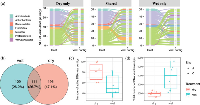
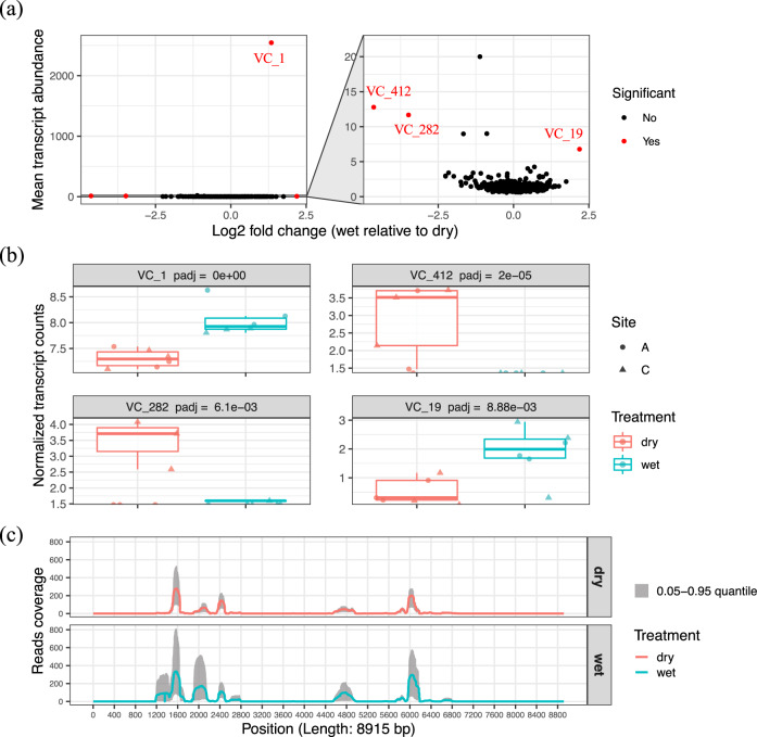
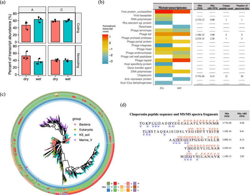
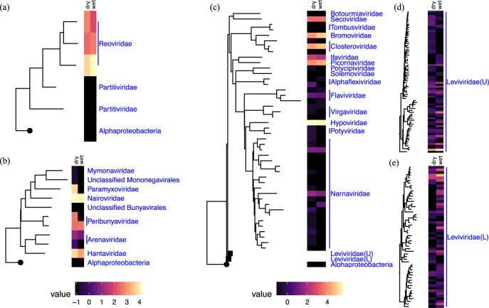
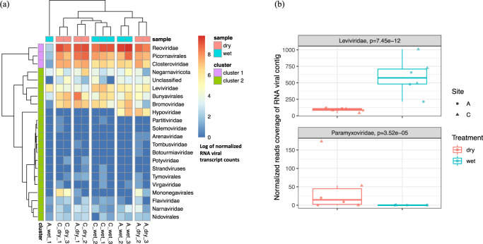
Soil is known to harbor viruses, but the majority are uncharacterized and their responses to environmental changes are unknown. Here, we used a multi-omics approach (metagenomics, metatranscriptomics and metaproteomics) to detect active DNA viruses and RNA viruses in a native prairie soil and to determine their responses to extremes in soil moisture. The majority of transcribed DNA viruses were bacteriophage, but some were assigned to eukaryotic hosts, mainly insects. We also demonstrated that higher soil moisture increased transcription of a subset of DNA viruses. Metaproteome data validated that the specific viral transcripts were translated into proteins, including chaperonins known to be essential for virion replication and assembly. The soil viral chaperonins were phylogenetically distinct from previously described marine viral chaperonins. The soil also had a high abundance of RNA viruses, with highest representation of Reoviridae. Leviviridae were the most diverse RNA viruses in the samples, with higher amounts in wet soil. This study demonstrates that extreme shifts in soil moisture have dramatic impacts on the composition, activity and potential functions of both DNA and RNA soil viruses.


Conflict of interest statement

The authors declare no competing interests.

Figures

Fig. 1
Fig. 1. Transcribed DNA viral communities and their responses to wet and dry soil conditions.
a An alluvium plot that illustrates pairings of the transcribed DNA viral contigs to putative host phyla. The transcribed DNA viral community was comprised of viral contigs from the curated DNA viral databases that were mapped by quality-filtered metatranscriptomic reads. The alluvia are colored by host taxa (first x axis of each sub-panel) assigned to respective transcribed DNA viral contigs (second x axis of each sub-panel). b A Venn diagram showing the number of unique transcribed DNA viral contigs detected in both wet and dry soils and ones exclusively detected in one of the soils. c Number of unique DNA viral contigs detected. A t-Test shows significantly more DNA contigs were transcribed in dry soil (p = 0.044). d Number of transcripts that mapped to the DNA viral contigs. For panels (c) and (d), the two independent field sites of Konza Experimental Field Station are indicated as site A (circles) and site C (triangles), with the wet soil in blue and dry soil in red.
Fig. 2
Fig. 2. DNA viral contigs with differential transcription in wet and dry soil treatments.
a Transcript abundance profiling of the identified DNA viral contigs. The mean transcript abundance of each DNA viral contig detected in all soils was plotted along the y axis in the sub-panel on the left. The normalized transcript abundances of each DNA viral contig were compared across treatments (wet and dry soils) and transformed into log2 fold change (wet relative to dry). The viral contigs with significantly differential transcript counts across treatments (p < 0.05) are highlighted in red in the sub-panel on the left. A zoomed-in panel on the right shows the viral contigs with lower transcript counts. b Four DNA viral contigs that were detected with differential transcript abundances in wet (blue) and dry (red) soils are shown. c Quality-filtered metatranscriptomic read coverage for the VC_1 sequence (total length of 8915 bp); the sequence with the highest number of transcripts mapped in (a). The solid line represents the mean read coverage per position detected in all replicates for each treatment (red = dry soil; blue = wet soil). The gray shading shows the range of read coverage distribution per position (0.05–0.95 quantile).
Fig. 3
Fig. 3. Functional characterization of viral transcripts and proteins.
a The percentage of the quality-filtered transcripts that mapped to gene-coding and noncoding regions of DNA viral contigs. The percentage of noncoding transcripts trended towards higher, but not significant, levels in drier soils at site A. b Counts of viral structural/functional groups that were detected in both the metatranscriptomes (heatmap on the left) and metaproteomes (table on the right). c A phylogenetic tree based on the protein alignment of bacterial (red), eukaryotic (green), and soil (blue)/marine (purple) viral chaperonins (GroEL-like). The soil viral chaperonin protein sequences were translated from the predicted genes in transcribed DNA viral contigs. An example of a conserved region (position 1–6 of the trimmed multiple sequence alignments in Supplementary Data 6) is shown in a six-track ring outside the tree and the six tracks represent the six amino acids from that region in order from the inner to the outer rings. The corresponding amino acid of the conserved region in each chaperonin sequence is color-coded and specified in the figure key. d Examples of highly confident viral chaperonin peptide sequences with their observed fragmentation ions (blue for b-ions and red for y-ions) in MS/MS spectra, along with their minimal peptide-spectrum match (PSM) scores and minimal mass error of precursor ions (PPM) from MSGF+ search results.
Fig. 4
Fig. 4. Phylogenetic diversity of RNA viruses and their estimated abundances.
Phylogenetic placement and abundance estimates of the detected: a double-stranded RNA viruses; b negative single-stranded RNA viruses; and ce positive single-stranded RNA viruses. Each of the RNA viral phylogenetic trees was constructed based on the aligned RNA-dependent RNA polymerase (RdRP) genes assigned to the identified RNA viral contigs and re-rooted by an RNA-dependent DNA polymerase (APO57079.1) of an Alphaproteobacterium (circular tree node). The abundance estimates for each taxon shown on the tree were measured by taking the average of the estimated average base-coverage per RNA viral contig mapping to this cluster of viruses detected under ‘dry’ or ‘wet’ conditions (left and right sections of each heatmap, respectively). The abundance estimates for each condition were then log transformed and illustrated in the heatmaps that are aligned to the respective tree tips. Two Leviviridae clades are collapsed in panel (c) for ease of visualization (tree nodes in rectangles); upper clade is noted as Leviviridae(U) and lower clade as Leviviridae(L). The phylogenetic structures and the abundance estimates for Leviviridae(U) and Leviviridae(L) are shown in panels (d) and (e), respectively.
Fig. 5
Fig. 5. Composition profiling of the RNA viral community and its abundance shift in response to wet and dry soil treatments.
a The RNA viral abundance of each phylogenetic group across all soil samples were summarized and clustered by composition similarity. b Two phylogenetic groups were detected with differential abundances in soils with different moistures (site A, circles; site C, triangles). Significantly more Leviviridae and significantly fewer Paramyxoviridae were detected in wet soils (blue) compared to dry (red) soils (p < 0.05).

References

    1. Paez-Espino D, et al. Uncovering Earth’s virome. Nature. 2016;536:425. doi: 10.1038/nature19094. - DOI - PubMed
    1. Emerson JB, et al. Host-linked soil viral ecology along a permafrost thaw gradient. Nat. Microbiol. 2018;3:870. doi: 10.1038/s41564-018-0190-y. - DOI - PMC - PubMed
    1. Starr EP, Nuccio EE, Pett-Ridge J, Banfield JF, Firestone MK. Metatranscriptomic reconstruction reveals RNA viruses with the potential to shape carbon cycling in soil. Proc. Natl Acad. Sci. USA. 2019;116:25900–25908. doi: 10.1073/pnas.1908291116. - DOI - PMC - PubMed
    1. Trubl G, et al. Soil viruses are underexplored players in ecosystem carbon processing. mSystems. 2018;3:e00076–00018. doi: 10.1128/mSystems.00076-18. - DOI - PMC - PubMed
    1. Schulz F, et al. Hidden diversity of soil giant viruses. Nat. Commun. 2018;9:4881. doi: 10.1038/s41467-018-07335-2. - DOI - PMC - PubMed

Publication types