Review
. 2021 Sep;18(9):1013-1026.
doi: 10.1038/s41592-021-01206-3. Epub 2021 Aug 26.

The power of imaging to understand extracellular vesicle biology in vivo

Affiliations
Review

The power of imaging to understand extracellular vesicle biology in vivo

Frederik J Verweij et al. Nat Methods. 2021 Sep.

Abstract

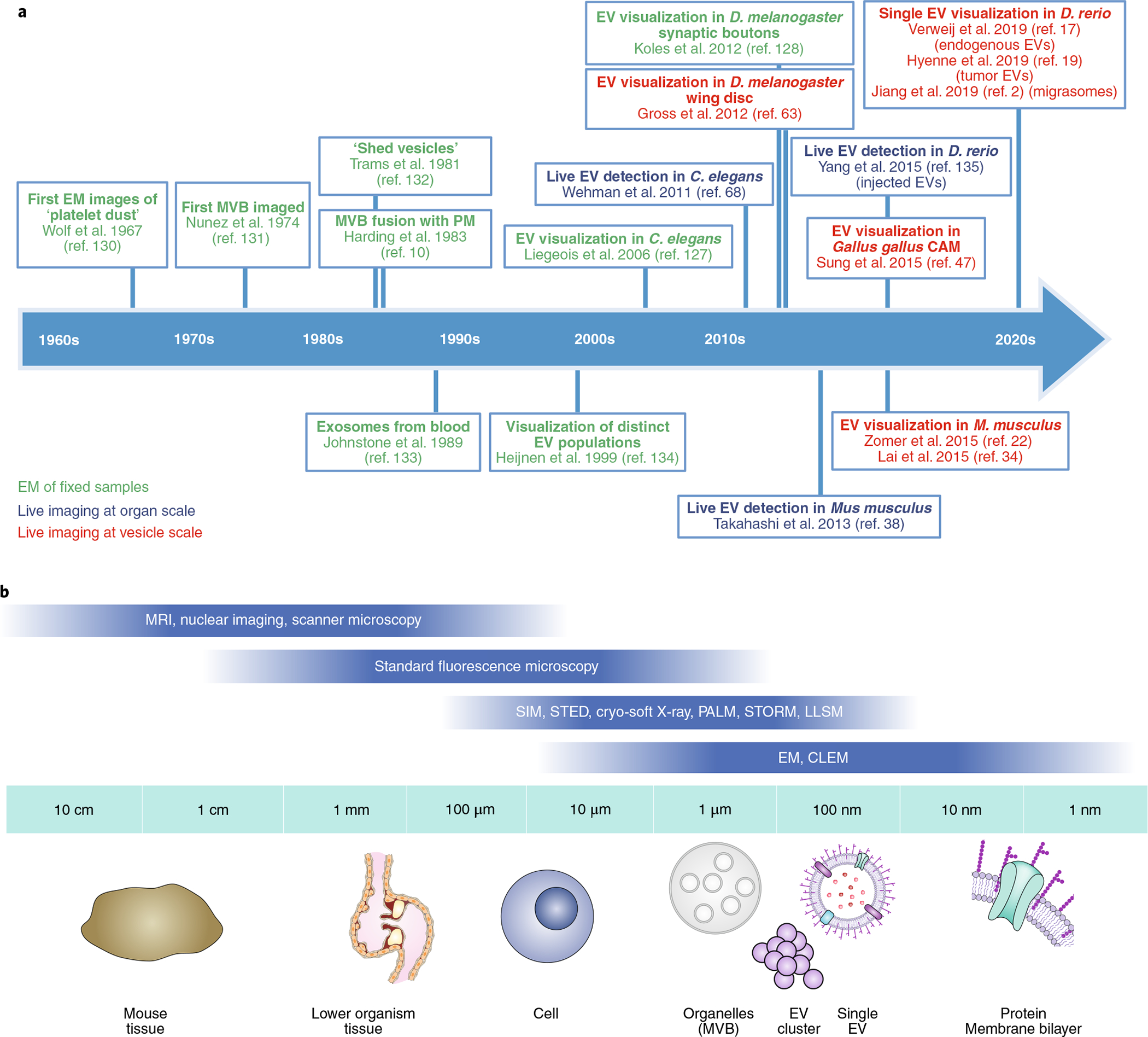
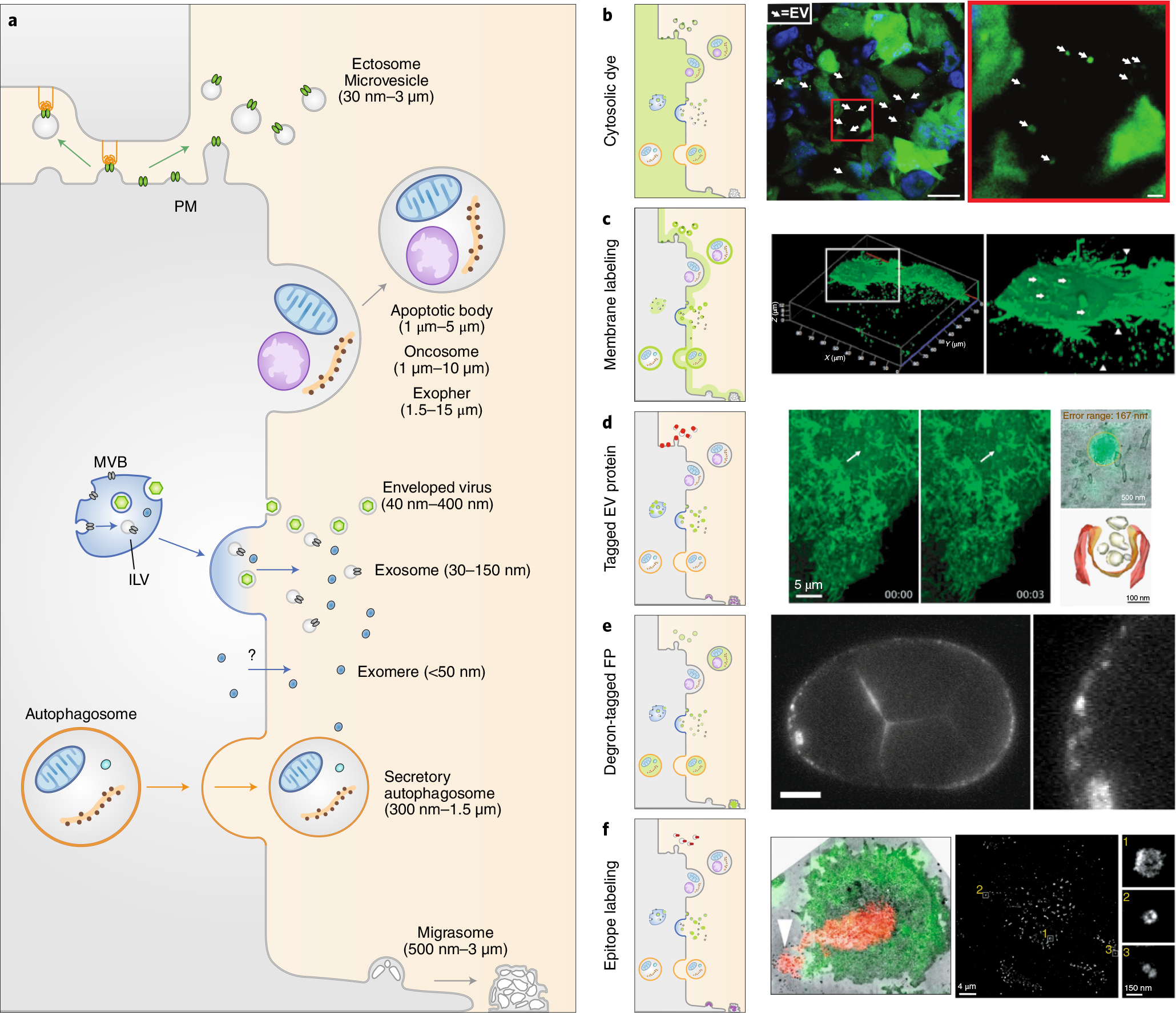
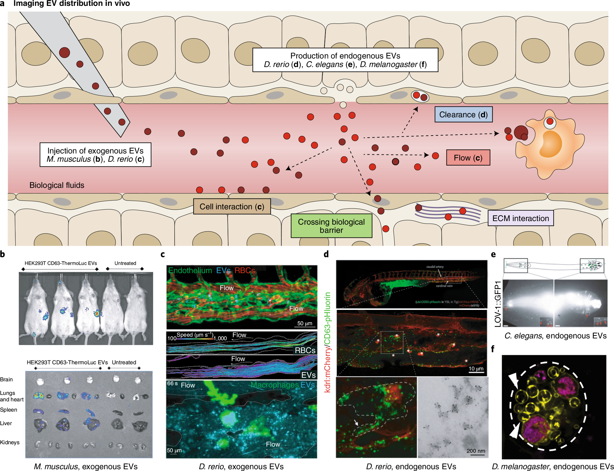
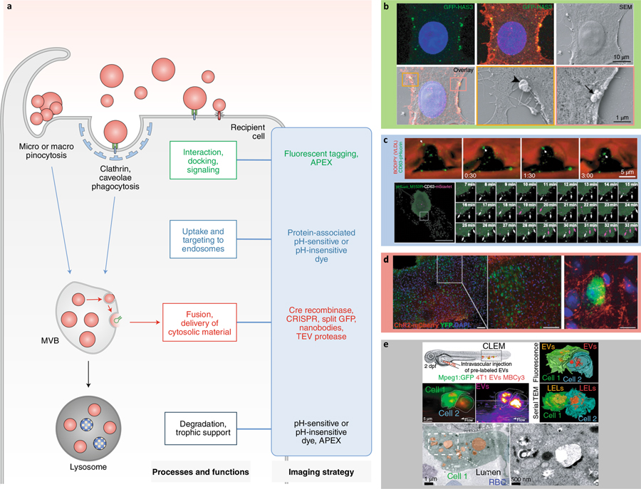
Extracellular vesicles (EVs) are nano-sized lipid bilayer vesicles released by virtually every cell type. EVs have diverse biological activities, ranging from roles in development and homeostasis to cancer progression, which has spurred the development of EVs as disease biomarkers and drug nanovehicles. Owing to the small size of EVs, however, most studies have relied on isolation and biochemical analysis of bulk EVs separated from biofluids. Although informative, these approaches do not capture the dynamics of EV release, biodistribution, and other contributions to pathophysiology. Recent advances in live and high-resolution microscopy techniques, combined with innovative EV labeling strategies and reporter systems, provide new tools to study EVs in vivo in their physiological environment and at the single-vesicle level. Here we critically review the latest advances and challenges in EV imaging, and identify urgent, outstanding questions in our quest to unravel EV biology and therapeutic applications.


Conflict of interest statement

Competing interests

D.R.F.C. is employed by Evox Therapeutics. S.E.A. serves on the Scientific Advisory Board of EVOX Therapeutics. All other authors have no competing interests.

Figures

Fig. 1 |
Fig. 1 |. Timeline of EV imaging milestones and broad overview of microscopy techniques to resolve EVs at different scales.
a, Timeline of imaging milestones in EV research. EM, electron microscopy. Refs. ,,,,,,,,,,,,–. b, Schematic of the resolution range of different microscopic approaches to resolve EVs at increasing resolution.
Fig. 2 |
Fig. 2 |. Tagging strategies to image EV production.
a, EVs are diverse double-leaflet membrane-enclosed structures generated from the PM (microvesicles, apoptotic bodies, oncosomes, exophers, enveloped viruses, and migrasomes), from endosomal compartments (exosomes and enveloped retroviruses), and from autophagic compartments (secretory autophagosomes). The origin of exomeres is still uncertain. bd, Tagging strategies to image EVs. Cytoplasmic labeling facilitates pan-EV tagging by labeling the cell cytosol and the lumen of any EVs (b). Right, large EVs released from MDA-MB-231 cells expressing Dendra2 in mice mammary glands. Arrows, EVs. Scale bars: 10 mm (left image), 1 mm (right image). Membrane labeling tags multiple EV subtypes (c). Right, confocal microscopy of live PalmGFP-expressing 293 T cells releasing EVs. Arrows, bud-like structure from the surface; arrowheads, processes extending from cells. Expressing tagged cargo proteins allows the tracking of EV subtypes (d). Right, live imaging of a burst of CD63-pHluorin fluorescence at the HeLa cell surface (arrows, fusion event), overlaid using CLEM (top right image) to observe an MVB fusing with the PM to release exosomes (bottom right image). e, Expression of degron-tagged fluorescent proteins allows EV tagging while cytosolic fluorescence in the source cell is degraded. Right, PH::CTPD-labeled EVs released from the unlabeled PM in C. elegans. Scale bar, 10 μm. f, Targeting of EV surface proteins by antibodies. Right, optical-EM correlation of M. musculus T cell that released EVs (red). Arrowhead, released microvesicles. Single EV imaging by dSTORM analysis of antibody staining (right image and insets).
Fig. 3 |
Fig. 3 |. Imaging EV propagation in vivo.
a, EV biodistribution can be mapped in the complex architecture of an organism after injection of labeled exogenous EVs or tagging endogenous EVs in situ. The in vivo fates of EVs (white boxes) are shown. bd, Imaging using injected or endogenous EVs in live animals. b, EV accumulation tracked at the organ scale using CD63-ThermoLuciferase in mice. c, EVs interacting with endothelial cells (green, top) or macrophages (green, bottom) tracked live in transparent zebrafish D. rerio; EV circulation in comparison to red blood cells (RBCs, red, middle). d, Endogenous EV clearance by scavenger endothelial cells and macrophages in D. rerio (top panels); inset of macrophage internalizing EVs captured from blood flow by SEC (bottom left); and IEM confirms the vesicular nature of the CD63-pHluorin signal in situ (bottom right). Arrow, macrophage protrusion; asterisks, macrophages. e, Fluorescently tagged EV cargo proteins track released EVs in C. elegans. Red arrows, EVs surrounding the head and tail. Scale bars, 10 μm. f, Fluorescently tagged EV cargo proteins track EVs released from giant secretory MVB-like compartments in D. melanogaster.
Fig. 4 |
Fig. 4 |. Tagging strategies to image EV interaction, uptake, and fate.
a, Different tagging strategies (blue box) reveal distinct aspects of EV–cell interactions. TEV, tobacco etch virus. be, Imaging strategies to track the fate and functions of EVs. b, Correlative light and scanning electron microscopy shows GFP-HAS3-labeled EVs interacting with the PM of receiving cells. Arrowhead, large EV; arrow, cluster and individual EVs of different sizes. c, Tracking uptake of endogenous CD63-pHluorin-labeled EVs in the D. rerio vasculature (top); and tracking double-labeled pHluorin-CD63-mScarlet EVs inside and outside HT1080 cells (bottom). VLDL, very low density lipoprotein. Arrows, internalized EV (top). White arrows, external EVs; purple arrows, internal EVs (bottom). d, Ex vivo mapping of EV mRNA using a Cre recombinase strategy in the mouse brain. e, Correlative light and electron microscopy shows Membright Cy3 lipid dye-labeled EVs accumulating in endolysosomes in D. rerio patrolling macrophages. dpf, days post-fertilization; LEL, late endo-lysosome; RBC, red blood cell.

References

    1. Van Niel G, D’Angelo G & Raposo G Shedding light on the cell biology of extracellular vesicles. Nat. Rev. Mol. Cell Biol 19, 213–228 (2018). - PubMed
    1. Jiang D et al. Migrasomes provide regional cues for organ morphogenesis during zebrafish gastrulation. Nat. Cell Biol 21, 966–977 (2019). - PubMed
    1. Huang Y et al. Migrasome formation is mediated by assembly of micron-scale tetraspanin macrodomains. Nat. Cell Biol 21, 991–1002 (2019). - PubMed
    1. Nicolás-Ávila JA et al. A network of macrophages supports mitochondrial homeostasis in the heart. Cell 183, 94–109.e23 (2020). - PubMed
    1. Melentijevic I et al. C. elegans neurons jettison protein aggregates and mitochondria under neurotoxic stress. Nature 542, 367–371 (2017). - PMC - PubMed

Publication types