Review
. 2021 Aug 10:12:722889.
doi: 10.3389/fphar.2021.722889. eCollection 2021.

An Overview of Cell-Based Assay Platforms for the Solute Carrier Family of Transporters

Affiliations
Review

An Overview of Cell-Based Assay Platforms for the Solute Carrier Family of Transporters

Vojtech Dvorak et al. Front Pharmacol. .

Abstract

The solute carrier (SLC) superfamily represents the biggest family of transporters with important roles in health and disease. Despite being attractive and druggable targets, the majority of SLCs remains understudied. One major hurdle in research on SLCs is the lack of tools, such as cell-based assays to investigate their biological role and for drug discovery. Another challenge is the disperse and anecdotal information on assay strategies that are suitable for SLCs. This review provides a comprehensive overview of state-of-the-art cellular assay technologies for SLC research and discusses relevant SLC characteristics enabling the choice of an optimal assay technology. The Innovative Medicines Initiative consortium RESOLUTE intends to accelerate research on SLCs by providing the scientific community with high-quality reagents, assay technologies and data sets, and to ultimately unlock SLCs for drug discovery.

Keywords: SLC; cell-based assay; chemical screening; drug discovery; solute carrier; transporters.


Conflict of interest statement

Research of the RESOLUTE consortium is in the precompetitive space. Authors PA, FD, AS, HS, MW are employed by Vifor. HB and EB are employed by Bayer. DZ was employed by Bayer during the manuscript preparation. TL and FB are employed by Sanofi. RR, FS, LS and ST are employed by Axxam. CS is employed by Pfizer. The remaining authors declare that the research was conducted in the absence of any commercial or financial relationships that could be constructed as a potential conflict of interest.

Figures
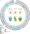
FIGURE 1
FIGURE 1
Solute carrier transporters, biochemical properties. (A) Schematic representation of the biochemical features of all SLC transporters. The superfamily is divided in 66 canonical sub-families and 5 non-canonical sub-families. For each SLC, the localization at the plasma membrane, the electrogenicity and the main substrate class are annotated. Annotation information regarding localization and substrate was extracted from Meixner et al. (2020) (updated by addition of SLC66 family), information regarding electrogenicity is referenced in Supplementary Table S1 and SLC fold was extracted from the Pfam database. (B) Transport mechanisms of SLCs. (C) Different association states are displayed by functional SLCs. PDB IDs 4ZW9, 6IRT and 6RVX were processed using Illustrate (Goodsell et al., 2019) to generate the visual representations.
FIGURE 2
FIGURE 2
Overview of the types of cell-based transport assays described in this review. Uptake assays directly measure the changes in the transported substrate across a cellular membrane. Binding assays report on protein stabilization upon binding of a molecule to the SLC in a cellular environment. Functional assays assess secondary effects in cells as a consequence of substrate transport.
FIGURE 3
FIGURE 3
Transport assay using a genetically encoded biosensor. The exemplified assay uses a protein sensor to detect changes in cellular pH caused by the substrate transported by the SLC. The sensor encodes a pH sensitive green fluorescent protein (GFP) linked to a red fluorescent protein (RFP – used for normalization). Wild-type (WT) cells have a neutral cytoplasmic pH where GFP is active. Upon overexpression of SLC9B2 (a proton importer) and addition of its substrate, the increased concentration of protons lowers the cytoplasmic pH. This causes the quenching of the GFP and therefore a decrease in fluorescence intensity compared to WT cells.
FIGURE 4
FIGURE 4
Schematic view of the MS-based transport assay for SLCs. Cells are incubated in medium or plasma containing a mix of metabolites, drugs and ions. After incubation, medium and/or intracellular fractions are extracted and prepared for MS analysis, followed by alignment and identification of molecules or ions. Both the comparison of identified molecules or ions in cellular extracts and medium as well as the comparison of cells with the SLC of interest knocked-out and overexpressed enable the identification of the metabolites, drugs or ions that are transported by the SLC of interest.
FIGURE 5
FIGURE 5
Cellular binding assay based on thermal shift. Cells are incubated with the molecule of interest, lysed, and exposed to increasing temperature. The remaining protein in native conformation is quantified by western blotting or using reporters. Binding of a small molecule stabilizes the protein of interest and leads to a shift in the melting temperature of the protein of interest.
FIGURE 6
FIGURE 6
Transport assay using a membrane potential dye. This assay uses a chemical dye to detect changes in the membrane potential (MP) caused by the ions transported by an electrogenic SLC. The dye coupled to a quencher is added to the medium. In the resting state, some dye enters the cell causing a fluorescent intensity that serves as a reference. Upon membrane hyperpolarization the dye does not penetrate in the cells and remains attached to the quencher, resulting in a fluorescence decrease. Upon depolarization of the membrane the dye detaches from the quencher and penetrates into the cells, eliciting a signal increase. Overexpression of SLC4A4 (a 1:Na+/3:HCO3− co-transporter) and addition of its substrates leads to hyperpolarization and a decrease in fluorescence intensity over time compared to wild-type cells.
FIGURE 7
FIGURE 7
SSM-based electrophysiology applied to SLCs. Membrane preparations from cells overexpressing the SLC of interest are applied to the sensor and together form a capacitively coupled membrane system. Therefore, charge translocation at the protein containing membrane can be detected via the SSM. After addition of the SLC substrate, changes in membrane potential are recorded. Only transient currents are measured, and the peak current represents the maximum speed of the transport.
FIGURE 8
FIGURE 8
SLC-GPCR coupling assay applied to SLC63A2. Sphingosine is phosphorylated by Sphk1/2 and exported by SLC63A2 OE cells through SLC63A2 into medium. Supernatant from these cells is then applied to detector cells, which stably express a S1P specific GPCR and the Ca2+ reporter Obelin. Activation of the GPCR as a surrogate readout for SLC63A2 transport of S1P is quantified by the increase of reporter fluorescence.
FIGURE 9
FIGURE 9
TRACT assay. Activation of a GPCR leads to changes in cellular morphology which can be quantified using the xCELLigence system. Exogenous addition of a SLC substrate which is at the same time a GPCR ligand to cells expressing both the SLC and the GPCR will lead to partial uptake and activation of the GPCR, measured by morphological changes with the xCELLigence real-time cell analysis (RTCA) instrument. Overexpression of the SLC leads to increased uptake of the substrate, which attenuates the GPCR-mediated cell response. When SLC transport is blocked by an inhibitor, the extracellular concentration of the SLC substrate/GPCR ligand is increased which leads to augmented activation of the GPCR and an enhanced cell response.
FIGURE 10
FIGURE 10
SLC coupling to nuclear hormone receptor applied to SLC10A6. A cell line expressing a reporter plasmid combining a response element of the estrogen receptor alpha (ERα) and a luciferase encoding gene is treated with estrone sulfate, which is imported to the cytoplasm by SLC10A6 and cleaved by the steroid sulfatase. The product estrone then binds to the estrone-responsive element and activates luciferase expression from the reporter. Inducible overexpression of SLC10A6 leads to increased uptake of estrone sulfate and therefore increased luciferase intensity.
FIGURE 11
FIGURE 11
Phenotypic assay based on synthetic lethality. WT cells are expressing SLCA and SLCB which are two transporters with a strong negative genetic interaction. KO of SLCA results in cells dependent on SLCB and vice versa. Therefore, selective inhibitors of SLCA kill only SLCB KO cells.
FIGURE 12
FIGURE 12
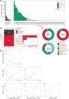
Overview and comparison of assay techniques in use. (A) Number of assays reported in ChEMBL per SLC family. (B) Distribution of assays based on assay format and assay type (cell-based assays only). Assay format was determined from the BAO label reported in the ChEMBL database (e.g. cell-based format and single protein format). Assay type was assigned according to manually created rules (see supplementary material) (C) Detection methods employed by each assay type (cell-based assays only). The detection method was assigned to the different categories based on manually created rules (see Supplementary table 2) (D) Comparison of IC50 values of SLC13A5 inhibitors obtained by different assays (Data retrieved from Huard et al. (2016); Pajor et al. (2016)); IC50 of >10, or >30 µM respectively, refers to the detection limit).

References

    1. Abu-Remaileh M., Wyant G. A., Kim C., Laqtom N. N., Abbasi M., Chan S. H., et al. (2017). Lysosomal Metabolomics Reveals V-ATPase- and mTOR-dependent Regulation of Amino Acid Efflux from Lysosomes. Science 358, 807–813. 10.1126/science.aan6298 - DOI - PMC - PubMed
    1. Adelmann C. H., Traunbauer A. K., Chen B., Condon K. J., Chan S. H., Kunchok T., et al. (2020). MFSD12 Mediates the Import of Cysteine into Melanosomes and Lysosomes. Nature 588, 699–704. 10.1038/s41586-020-2937-x - DOI - PMC - PubMed
    1. Al-Khawaja A., Haugaard A. S., Marek A., Löffler R., Thiesen L., Santiveri M., et al. (2018). Pharmacological Characterization of [3H] ATPCA as a Substrate for Studying the Functional Role of the Betaine/GABA Transporter 1 and the Creatine Transporter. ACS Chem. Neurosci. 9, 545–554. 10.1021/acschemneuro.7b00351 - DOI - PubMed
    1. Al-Khawaja A., Petersen J. G., Damgaard M., Jensen M. H., Vogensen S. B., Lie M. E. K., et al. (2014). Pharmacological Identification of a Guanidine-Containing β-Alanine Analogue with Low Micromolar Potency and Selectivity for the Betaine/GABA Transporter 1 (BGT1). Neurochem. Res. 39, 1988–1996. 10.1007/s11064-014-1336-9 - DOI - PubMed
    1. Alexandrov A. I., Mileni M., Chien E. Y. T., Hanson M. A., Stevens R. C. (2008). Microscale Fluorescent Thermal Stability Assay for Membrane Proteins. Structure 16, 351–359. 10.1016/j.str.2008.02.004 - DOI - PubMed

LinkOut - more resources