. 2021 Aug 10:12:616215.
doi: 10.3389/fimmu.2021.616215. eCollection 2021.

Oral Fc-Coupled Preproinsulin Achieves Systemic and Thymic Delivery Through the Neonatal Fc Receptor and Partially Delays Autoimmune Diabetes

Affiliations

Oral Fc-Coupled Preproinsulin Achieves Systemic and Thymic Delivery Through the Neonatal Fc Receptor and Partially Delays Autoimmune Diabetes

Noémie Corcos et al. Front Immunol. .

Abstract

Tolerogenic vaccinations using beta-cell antigens are attractive for type 1 diabetes prevention, but clinical trials have been disappointing. This is probably due to the late timing of intervention, when multiple auto-antibodies are already present. We therefore devised a strategy to introduce the initiating antigen preproinsulin (PPI) during neonatal life, when autoimmunity is still silent and central tolerance mechanisms, which remain therapeutically unexploited, are more active. This strategy employs an oral administration of PPI-Fc, i.e. PPI fused with an IgG Fc to bind the intestinal neonatal Fc receptor (FcRn) that physiologically delivers maternal antibodies to the offspring during breastfeeding. Neonatal oral PPI-Fc vaccination did not prevent diabetes development in PPI T-cell receptor-transgenic G9C8.NOD mice. However, PPI-Fc was efficiently transferred through the intestinal epithelium in an Fc- and FcRn-dependent manner, was taken up by antigen presenting cells, and reached the spleen and thymus. Although not statistically significant, neonatal oral PPI-Fc vaccination delayed diabetes onset in polyclonal Ins2-/-.NOD mice that spontaneously develop accelerated diabetes. Thus, this strategy shows promise in terms of systemic and thymic antigen delivery via the intestinal FcRn pathway, but the current PPI-Fc formulation/regimen requires further improvements to achieve diabetes prevention.

Keywords: T cells; autoimmunity; immune tolerance; neonatal Fc receptor (FcRn); preproinsulin; thymus; type 1 diabetes; vaccination.


Conflict of interest statement

The authors declare that the research was conducted in the absence of any commercial or financial relationships that could be construed as a potential conflict of interest.

Figures

Figure 1
Figure 1
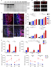
Oral PPI-Fc does not prevent diabetes in TCR-transgenic G9C8.NOD mice. (A) Diabetes incidence in G9C8.NOD mice force-fed at day 1 with 50 µg PPI-Fc (red) or equimolar amounts of PPI (blue), OVA-Fc (green) or PBS (black). Diabetes was induced by prime-boost immunization with PIB15–23 peptide and CpG at 4 and 6 weeks of age. (B–E) Proportion of CD8+ and CD4+ T cells out of total TCR+ cells (top panels) and of Foxp3 and Foxp3+ T cells out of total CD4+ T cells (bottom panels) in the spleen (B), PLNs (C) and MLNs (D) of 4-week-old PPI-Fc- or control-treated G9C8.NOD mice. (E) Splenocytes from 4-week-old PPI-Fc- or control-treated G9C8.NOD mice were stimulated in vitro with increasing concentrations of PPIB15-23 peptide for 5 h and assessed for surface CD69 and CD107a expression and production of IFN-γ and TNF-α. Results are from 4-5 separate experiments with 7-9 mice/group; bars indicate mean ± SEM values.
Figure 2
Figure 2
Oral PPI-Fc crosses the gut epithelium in an FcRn-dependent manner and is taken up by LP DCs and macrophages. (A) Age-dependent FcRn expression in the gut epithelium. Intestinal epithelial cells were isolated from the whole digestive tract of 1-day-old to 13-week-old G9C8.NOD mice and 1-day-old FcRn-/-.B6 mice and probed for FcRn, and Hsp70 as loading control. (B) One-day-old wild-type G9C8.NOD (WT) or FcRn-/-.B6 mice were force-fed with 50 µg of AF680-labeled PPI-Fc. Ex-vivo imaging of the intestines of WT and FcRn-/- mice (n=2/each) 4, 24 or 48 h after PPI-Fc force-feeding. (C) Confocal images of the same intestines recovered from WT and FcRn-/-.B6 mice at 24 h (20X and 40X magnifications). Sections were stained for insulin (white), CD11c (green) and EpCAM (red), with nuclei counterstained with DAPI (blue). (D) Five-day-old G9C8.NOD mice were force-fed with 50 µg AF647-labeled PPI-Fc (red bars), PPI (blue), or PBS (black). LP cells were isolated 4 or 8 h after gavage, and protein uptake (AF647 median fluorescence) was analyzed in CD103+ and CD103 cDCs and CX3CR1+CD11b and CX3CR1CD11b+ macrophages (MΦ). (E) Five-day-old G9C8.NOD mice were force-fed with 50 µg AF647-labeled IgG1 (red), F(ab’)2 (blue), or PBS (black). Four hours later, protein uptake was analyzed in the LP for the indicated APC subsets: CD103+ and SIRPα+ cDCs (left) and CX3XR1+, CX3CR1SIRPαlo and CX3CR1SIRPαhi macrophages (MΦ, right). (F) Comparison of the AF647 median fluorescence at 4 and 24 h in the same APC subsets. Results in (D–F) are expressed as mean ± SEM from 3 mice per group. *P ≤ 0.03, **P ≤ 0.007, ***P ≤ 0.0002 by 2-way ANOVA.
Figure 3
Figure 3
APC subsets in the neonatal LP and spleen. Cells were gated on live CD45+LinB220 events (see Supplementary Figures 3, 4 for details). (A) In the LP, CD11chi cDCs (top left panel) are divided into a CD103+ and a SIRPα+ subset (top right); F4/80+ macrophages are divided into a CX3CR1+CD11b (indicated as CX3CR1+) and a CX3CR1CD11b+ (indicated as CX3CR1) subset (bottom left). The latter comprises a SIRPαlo and SIRPαhi subpopulation (bottom right). (B) In the spleen, CD11chi cDCs are similarly composed of CD103+ and SIRPα+ subpopulations. F4/80+ macrophages are all CD11b+ and either CX3CR1+ or CX3CR1. The latter comprise a SIRPαlo and SIRPαhi subpopulation.
Figure 4
Figure 4
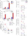
Oral PPI-Fc reaches the spleen mainly in soluble form and is taken up locally by SIRPα+ cDCs and macrophages. (A, B) Five-day-old G9C8.NOD mice were force-fed with 50 µg AF647-labeled IgG1 (red), F(ab’)2 (blue), or PBS (black). Four hours later, protein uptake (AF647 median fluorescence) was analyzed in the spleen for the indicated APC subsets: CD103+ and SIRPα+ cDCs (A) and CX3XR1+, CX3XR1SIRPαlo and CX3XR1SIRPαhi macrophages (B). (C) Comparison of the AF647 median fluorescence at 4 and 24 h in the same APC subsets. (D) One-day-old wild-type G9C8.NOD (WT) or FcRn-/-.B6 mice were force-fed with 50 µg of PPI-Fc (or PBS in WT mice), serum was collected after 4, 24 and 48 h and PPI-Fc concentrations measured by enzyme-linked immunosorbent assays. Results are expressed as mean ± SEM from 3 mice per group (6/group in D). *P ≤ 0.03, **P ≤ 0.006, ***P ≤ 0.0003 by 2-way ANOVA.
Figure 5
Figure 5
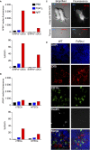
Oral PPI-Fc reaches the thymus mainly through ferrying by migratory SIRPαhi cDCs. (A, B) Five-day-old G9C8.NOD mice were force-fed with 50 µg AF647-labeled IgG1 (red), F(ab’)2 (blue), or PBS (black). Thymi from 3 mice/group were pooled and cells were magnetically enriched for CD11c+ DCs (A) and EpCAM+ TECs (B) before flow cytometry (see Supplementary Figure 5 for details). Protein uptake (AF647 median fluorescence and percentage of AF647+ cells) was analyzed in the indicated DC and TEC subsets. (C) Ex-vivo imaging of the whole body (top) and thymus (bottom) of 1-day-old G9C8.NOD mice 72 h after oral administration of 50 µg AF680-labeled PPI-Fc or PPI (day 4 of life). (D) Confocal images of thymic sections recovered from WT G9C8.NOD and FcRn-/-.B6 mice 24 h after oral administration of fluorescent PPI-Fc. Sections were stained for insulin (white), CD11c (green) and cytokeratin 5 (CK5, red), with nuclei counterstained with DAPI (blue).
Figure 6
Figure 6
Oral PPI-Fc partially delays diabetes onset in polyclonal Ins2 -/-.NOD mice. Diabetes incidence in Ins2 -/-.NOD mice after oral administration of 50 µg PPI-Fc (red) or equimolar amounts of PPI (blue), OVA-Fc (green), IgG1 (grey) or PBS (black) at day 1 (A) or day 1, 4, 7 and 10 of life (B; p=0.13 between PPI-Fc and control groups by log-rank test).

References

    1. James EA, Mallone R, Kent SC, DiLorenzo TP. T-Cell Epitopes and Neo-Epitopes in Type 1 Diabetes: A Comprehensive Update and Reappraisal. Diabetes (2020) 69:1311–35. 10.2337/dbi19-0022 - DOI - PMC - PubMed
    1. Mallone R, Eizirik DL. Presumption of Innocence for Beta Cells: Why are They Vulnerable Autoimmune Targets in Type 1 Diabetes? Diabetologia (2020) 63:1999–2006. 10.1007/s00125-020-05176-7 - DOI - PubMed
    1. Carré A, Richardson SJ, Larger E, Mallone R. Presumption of Guilt for T Cells in Type 1 Diabetes: Lead Culprits or Partners in Crime Depending on Age of Onset? Diabetologia (2021) 64:15–25. 10.1007/s00125-020-05298-y - DOI - PMC - PubMed
    1. Culina S, Boitard C, Mallone R. Antigen-Based Immune Therapeutics for Type 1 Diabetes: Magic Bullets or Ordinary Blanks? Clin Dev Immunol (2011) 2011:286248. 10.1155/2011/286248 - DOI - PMC - PubMed
    1. Brezar V, Carel JC, Boitard C, Mallone R. Beyond the Hormone: Insulin as an Autoimmune Target in Type 1 Diabetes. Endocr Rev (2011) 32:623–69. 10.1210/er.2011-0010 - DOI - PubMed

Publication types

MeSH terms

Grants and funding