Case Reports
. 2021 Aug 10:12:673453.
doi: 10.3389/fgene.2021.673453. eCollection 2021.

Case Report: A Novel Mutation in NFKB1 Associated With Pyoderma Gangrenosum

Affiliations
Case Reports

Case Report: A Novel Mutation in NFKB1 Associated With Pyoderma Gangrenosum

Ran Fang et al. Front Genet. .

Abstract

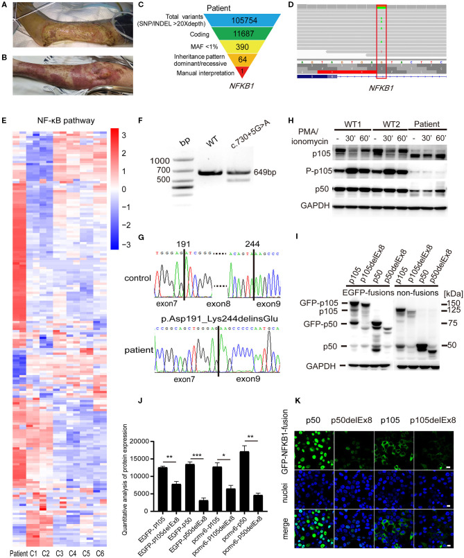
Pyoderma gangrenosum (PG) is a rare, destructive inflammatory skin disease of which a painful nodule or pustule breaks down to form a progressively enlarging ulcer. Ulcerations associated with PG may occur after trauma or injury to the skin. The etiology has not been clearly elucidated. Our report described a PG patient with a heterozygous splice-donor-site mutation in NFKB1 (c.730+5G>A) causing the absence of exon 8 and the formation of truncated p105 (p.Asp191_Lys244delinsGlu; p105delEx8), which led to distinct symptoms of high fever and excessive inflammation in wound area after routine surgical procedures. The functional analysis showed that the variant caused reduced phosphorylation of p105 and resulted in the decreased processing of p105 to p50. We conclude that the patient's symptoms were caused by dysregulation of the NF-κB signaling pathway.

Keywords: NF-κB signaling pathway; NFKB1; inflammation; novel mutation; pyoderma gangrenosum.


Conflict of interest statement

The authors declare that the research was conducted in the absence of any commercial or financial relationships that could be construed as a potential conflict of interest.

Figures

Figure 1
Figure 1
A pyoderma gangrenosum (PG) patient with heterozygous splice-donor-site mutation in NFKB1. (A) After the left knee joint replacement surgery, purple erythema appeared on the left leg of the patient, and a large area of ulcers gradually formed. (B) The patient's ulcer gradually improved after treatment with intravenous immunoglobulin (IVIG) plus glucocorticoid. (C) Schematic of the whole-exome sequencing (WES) data-filtering approach under the assumption of dominant/de novo inheritance, leading to the identification of an NFKB1 variant. For details of variants in each assumed inheritance, see Supplementary Tables 1, 2. INDEL, frameshift, or non-frameshift insertions and deletions; SNP, single-nucleotide polymorphisms including missense, splice-site, and stop-codon variants. (D) The integrative genomics viewer revealed the exome sequencing reads covering a heterozygous mutation (c.730+5G>A) in intron 8 splice donor site of NFKB1. (E) RNA-sequencing analysis of NF-κB target genes in patient's peripheral blood mononuclear cells (PBMCs) compared with those of six unaffected controls (C1–C6). Analysis of each sample was performed in duplicate. For gene names, see Supplementary Figure 1. (F) The expression of NFKB1 mRNA in PBMCs of healthy controls and the patient were analyzed by RT-PCR. Primers located in exons 6 and 11 were used to amplify exons 7–10 (649 bp) of NFKB1. In addition to the expected band, a shorter product was observed in the patient, suggesting that the mutation caused the deletion of an exon (exon 7, exon 8, or exon 9). (G) Sequencing of RT-PCR products from healthy control showed normal splicing (top). The patient's heterozygous mutation (c.730+5G>A) caused the in-frame skipping of exon 8 and the fusion of exon 7 and exon 9 (105delEx8, p.Asp191_Lys244delinsGlu) (bottom). (H) PBMCs from the patient and healthy controls were stimulated with phorbol 12-myristate 13-acetate (PMA) plus ionomycin. The amounts of p105 and p50 and the phosphorylation of p105 (Ser933, P-p105) were analyzed by Western blotting. GAPDH was used as the loading control. (I) HEK293T cells were transiently transfected with cytomegalovirus promoter-driven ectopic expression vector; the amount of ectopic protein was analyzed based on the results of Western blotting. As shown in the figure, on the left are expressions of fusion proteins wild-type and mutant p105 and p50, and on the right are non-fusion proteins. The 150-kDa band is GFP-p105. The 75-kDa band is GFP-p50. The 50-kDa faint band in all lanes is endogenous p50. The molecular weight marker is shown on the right. Only a weak fluorescent signal was detected for the mutant protein. (J) Grayscale analysis of the western blot results in (I). (K) The immunofluorescence results showed that GFP-p50 was localized in the nucleus and GFP-p105 was in the cytoplasm. Green, green fluorescent protein (GFP) fusion; blue, nuclear. The scale bar represents 10 μm. *P < 0.05, **P < 0.001, ***P < 0.0001.

References

    1. Alavi A., French L. E., Davis M. D., Brassard A., Kirsner R. S. (2017). Pyoderma gangrenosum: an update on pathophysiology, diagnosis and treatment. Am. J. Clin. Dermatol. 18, 355–372. 10.1007/s40257-017-0251-7 - DOI - PubMed
    1. Boztug H., Hirschmugl T., Holter W., Lakatos K., Kager L., Trapin D., et al. . (2016). NF-kappaB1 Haploinsufficiency causing immunodeficiency and EBV-Driven lymphoproliferation. J. Clin. Immunol. 36, 533–540. 10.1007/s10875-016-0306-1 - DOI - PMC - PubMed
    1. Braswell S. F., Kostopoulos T. C., Ortega-Loayza A. G. (2015). Pathophysiology of pyoderma gangrenosum (PG): an updated review. J. Am. Acad. Dermatol. 73, 691–698. 10.1016/j.jaad.2015.06.021 - DOI - PubMed
    1. Bryant V. L., Tangye S. G. (2016). The expanding spectrum of NFkB1 deficiency. J. Clin. Immunol. 36, 531–532. 10.1007/s10875-016-0310-5 - DOI - PubMed
    1. Defilippis E. M., Feldman S. R., Huang W. W. (2015). The genetics of pyoderma gangrenosum and implications for treatment: a systematic review. Br. J. Dermatol. 172, 1487–1497. 10.1111/bjd.13493 - DOI - PubMed

Publication types