

Protectin D1 protects against lipopolysaccharide-induced acute lung injury through inhibition of neutrophil infiltration and the formation of neutrophil extracellular traps in lung tissue
- PMID: 34447467
- PMCID: PMC8355679
- DOI: 10.3892/etm.2021.10508
Protectin D1 protects against lipopolysaccharide-induced acute lung injury through inhibition of neutrophil infiltration and the formation of neutrophil extracellular traps in lung tissue
Abstract
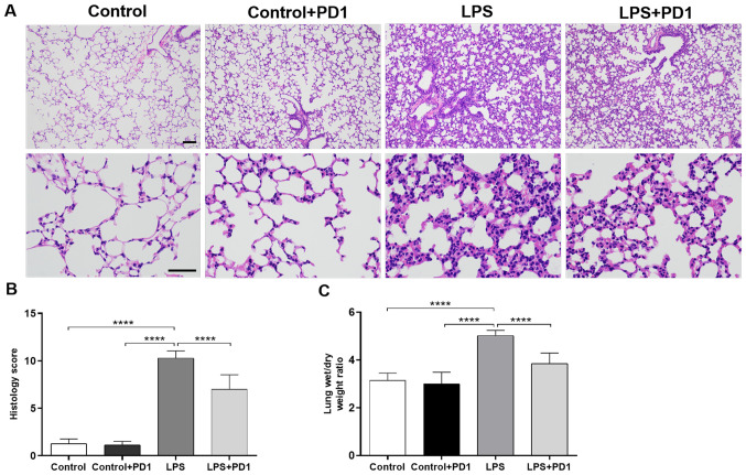
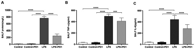
Protectin D1 (PD1), a DHA-derived lipid mediator, has recently been shown to possess anti-inflammatory and pro-resolving properties. To date, little is known about the effect of PD1 on lipopolysaccharide (LPS)-induced acute lung injury (ALI) in mice. The aim of the present study was to investigate the therapeutic effects of PD1 on LPS-induced ALI and its potential mechanisms of action. ALI was induced via an intraperitoneal injection of LPS, where PD1 (2 ng/mouse) was administered intravenously 30 min after LPS challenge. Mice were sacrificed 24 h after modeling. Lung histopathological changes were assessed using hematoxylin and eosin staining and myeloperoxidase (MPO) activity was tested using immunohistochemistry. Tumor necrosis-α and interleukin-6 levels in the bronchoalveolar lavage fluid (BALF) and serum were measured using ELISA. To detect neutrophil extracellular traps produced by infiltrated neutrophils in the lung tissue, immunofluorescence staining was performed using anti-MPO and anti-histone H3 antibodies. The results indicated that PD1 significantly attenuated histological damage and neutrophil infiltration in lung tissue, reduced the lung wet/dry weight ratio, protein concentration and proinflammatory cytokine levels in BALF and decreased proinflammatory cytokine levels in serum. Moreover, neutrophil citrullinated histone H3 (CitH3) expression was also reduced after PD1 administration. In conclusion, PD1 attenuated LPS-induced ALI in mice via inhibition of neutrophil extracellular trap formation in lung tissue. Therefore, PD1 administration may serve to be a new strategy for treating ALI.
Keywords: acute lung injury; neutrophil extracellular trap; neutrophil infiltration; protectin D1.
Copyright © 2020, Spandidos Publications.
Conflict of interest statement
The authors declare that they have no competing interests.
Figures


References
-
- Ferguson ND, Frutos-Vivar F, Esteban A, Fernández-Segoviano P, Aramburu JA, Nájera L, Stewart TE. Acute respiratory distress syndrome: Underrecognition by clinicians and diagnostic accuracy of three clinical definitions. Crit Care Med. 2005;33:2228–2234. doi: 10.1097/01.ccm.0000181529.08630.49. - DOI - PubMed
LinkOut - more resources
Full Text Sources
Research Materials
Miscellaneous