. 2021 Aug 10:8:697055.
doi: 10.3389/fmed.2021.697055. eCollection 2021.

Hip Position Acutely Affects Oxygenation and Perfusion of Kidney Grafts as Measured by Functional Magnetic Resonance Imaging Methods-The Bent Knee Study

Affiliations

Hip Position Acutely Affects Oxygenation and Perfusion of Kidney Grafts as Measured by Functional Magnetic Resonance Imaging Methods-The Bent Knee Study

Laila-Yasmin Mani et al. Front Med (Lausanne). .

Abstract

Background: Kidney perfusion and oxygenation are two important determinants of kidney graft function. In kidney transplantation, repeated graft hypoperfusion may occur during hip flexion, for example in the sitting position, due to the progressive development of fibrotic tissue around iliac arteries. The aim of this study was to assess the changes in oxygenation and perfusion of kidney grafts during hip flexion and extension using a new functional magnetic resonance imaging (fMRI) protocol. Methods: Nineteen kidney graft recipients prospectively underwent MRI on a 3T scanner including diffusion-weighted, blood oxygenation level dependent (BOLD), and arterial spin labeling sequences in hip positions 0° and >90° before and after intravenous administration of 20 mg furosemide. Results: Unexpectedly, graft perfusion values were significantly higher in flexed compared to neutral hip position. Main diffusion-derived parameters were not affected by hip position. BOLD-derived cortico-medullary R2* ratio was significantly modified during hip flexion suggesting an intrarenal redistribution of the oxygenation in favor of the medulla and to the detriment of the cortex. Furthermore, the increase in medullary oxygenation induced by furosemide was significantly blunted during hip flexion (p < 0.001). Conclusion: Hip flexion has an acute impact on perfusion and tissue oxygenation in kidney grafts. Whether these position-dependent changes affect the long-term function and outcome of kidney transplants needs further investigation.

Keywords: BOLD; arterial spin labeling; functional MRI; hip flexion; kidney transplantation; multiparametric magnetic resonance imaging; oxygenation; perfusion.


Conflict of interest statement

The authors declare that the research was conducted in the absence of any commercial or financial relationships that could be construed as a potential conflict of interest.

Figures

Figure 1
Figure 1
Study plan. Positioning order was alternated in each following subject, then maintained for the second part of measurements. eGFR, estimated glomerular filtration rate estimated according to chronic kidney disease epidemiology formula; AKI, acute kidney injury; BOLD, blood oxygen level-dependent magnetic resonance imaging; DWI, diffusion-weighted imaging; ASL, arterial spin labeling-magnetic resonance imaging; TOF-A, time of flight-angiography; *performed during hip flexion.
Figure 2
Figure 2
Flow chart. Patient screening and data exclusion. DWI, diffusion-weighted imaging; BOLD, blood oxygen level dependent-magnetic resonance imaging; ASL, arterial spin labeling-magnetic resonance imaging.
Figure 3
Figure 3
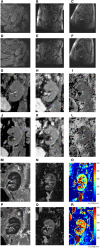
(A–R). Image examples (one subject). Anatomical sequence (T1 map) during neutral hip position (A–C) and hip flexion (D–F) in sagittal (A,D), coronal (B,E), and transverse (C,F) planes. Functional MRI sequences during neutral hip position (G–I, M–O) and hip flexion (J–L, P–R): total apparent diffusion coefficient (ADCT) map (G,J); pure diffusion coefficient (ADCD) map (H,K); fraction of perfusion (FP) map (I,L); resonance transverse relaxation rate (R2*) map (M,P); arterial spin labeling (ASL) map (N,O,Q,R).
Figure 4
Figure 4
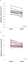
(A,B). Blood oxygen level dependent (BOLD)-MRI during neutral and flexed hip position. Ladder plots showing resonance transverse relaxation rate (R2*)-derived parameters during neutral and flexed hip position: (A) Medullary ratio R2* without/with (wo/w) furosemide (F), n = 13; (B) Medullary to cortical ratio R2*, n = 16. Paired Student's t-test-derived p-value comparing neutral and flexed hip position (p). Colors denote opposite trends.
Figure 5
Figure 5
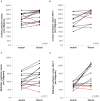
(A–D) Arterial spin labeling-MRI during neutral and flexed hip position. Ladder plots showing perfusion values measured in cortex (A,B) and medulla (C,D) without (A,C) and after (B,D) furosemide (F) administration during neutral and flexed hip position. (A) n = 16; (B) n = 11; (C) n = 15; (D) n = 13. Paired Student's t-test-derived p-value comparing neutral and flexed hip position (p). Colors denote opposite trends.

References

    1. Wolfe RA, Ashby VB, Milford EL, Ojo AO, Ettenger RE, Agodoa LY, et al. . Comparison of mortality in all patients on dialysis, patients on dialysis awaiting transplantation, and recipients of a first cadaveric transplant. N Engl J Med. (1999) 341:1725–30. 10.1056/NEJM199912023412303 - DOI - PubMed
    1. Meier-Kriesche HU, Schold JD, Srinivas TR, Kaplan B. Lack of improvement in renal allograft survival despite a marked decrease in acute rejection rates over the most recent era. Am J Transplant. (2004) 4:378–83. 10.1111/j.1600-6143.2004.00332.x - DOI - PubMed
    1. Pascual J, Perez-Saez MJ, Mir M, Crespo M. Chronic renal allograft injury: early detection, accurate diagnosis and management. Transplant Rev. (2012) 26:280–90. 10.1016/j.trre.2012.07.002 - DOI - PubMed
    1. Van Loon E, Senev A, Lerut E, Coemans M, Callemeyn J, Van Keer JM, et al. . Assessing the Complex Causes of Kidney Allograft Loss. Transplantation. (2020) 104:2557–66. 10.1097/TP.0000000000003192 - DOI - PubMed
    1. Kuss R, Teinturier J, Milliez P. [Some attempts at kidney transplantation in man]. Mem Acad Chir. (1951) 77:755–64. - PubMed