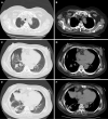
Nocardiosis with diffuse involvement of the pleura: A case report
- PMID: 34447831
- PMCID: PMC8362512
- DOI: 10.12998/wjcc.v9.i23.6824
Nocardiosis with diffuse involvement of the pleura: A case report
Abstract
Background: Nocardiosis is an uncommon infection that usually occurs in immunocompromised patients, and the pulmonary system is the most common site. We report an uncommon case of nocardiosis with diffuse involvement of the pleura, which presented as multiple localized nodular or hillock lesions on computed tomography (CT) with local chest wall infiltration.
Case summary: A 54-year-old woman was referred to our hospital due to cough and fever for 20 d. She had a history of nephrotic syndrome for 7 mo and was given prednisone (60 mg/d) 6 mo previously. The hormone was then gradually reduced to the current dose of 25 mg/d. Chest CT showed many nodular or hillock lesions in the right pleura, mediastinum, and interlobar fissure areas. On the lower layer, one lesion infiltrated the chest wall. She was treated with piperacillin sodium and sulbactam sodium, but the therapeutic effect was not good. In this regard, ultrasound-guided local infiltration anesthesia was further conducted for perihepatic hydrops drainage to improve diagnostic accuracy. Puncture fluid culture isolated Nocardia species, confirming the diagnosis of nocardiosis. Subtype Nocardia farcinica was identified by matrix-assisted laser desorption/ionization time-of-flight mass spectrometry. Antibiotic treatment was switched to trimethoprim/sulfamethoxazole and imipenem. After 8 d of treatment, the patient was discharged from the hospital with improved condition, and she has been recurrence-free for 2 years.
Conclusion: This report illustrates that nocardiosis should be suspected when clinicians encounter patients who are immunocompromised and have diffuse involvement of the pleura.
Keywords: Case report; Computed tomography; Lung; Nocardiosis; Pleura.
©The Author(s) 2021. Published by Baishideng Publishing Group Inc. All rights reserved.
Conflict of interest statement
Conflict-of-interest statement: The authors declare that they have no conflicts of interest.
Figures


References
-
- Restrepo A, Clark NM Infectious Diseases Community of Practice of the American Society of Transplantation. Nocardia infections in solid organ transplantation: Guidelines from the Infectious Diseases Community of Practice of the American Society of Transplantation. Clin Transplant. 2019;33:e13509. - PubMed
-
- Haussaire D, Fournier PE, Djiguiba K, Moal V, Legris T, Purgus R, Bismuth J, Elharrar X, Reynaud-Gaubert M, Vacher-Coponat H. Nocardiosis in the south of France over a 10-years period, 2004-2014. Int J Infect Dis. 2017;57:13–20. - PubMed
-
- Hui CH, Au VW, Rowland K, Slavotinek JP, Gordon DL. Pulmonary nocardiosis re-visited: experience of 35 patients at diagnosis. Respir Med. 2003;97:709–717. - PubMed
-
- Blackmon KN, Ravenel JG, Gomez JM, Ciolino J, Wray DW. Pulmonary nocardiosis: computed tomography features at diagnosis. J Thorac Imaging. 2011;26:224–229. - PubMed
Publication types
LinkOut - more resources
Full Text Sources

