. 2020 Sep;1(9):882-893.
doi: 10.1038/s43018-020-0095-6. Epub 2020 Aug 10.

Multifunctional oncolytic nanoparticles deliver self-replicating IL-12 RNA to eliminate established tumors and prime systemic immunity

Affiliations

Multifunctional oncolytic nanoparticles deliver self-replicating IL-12 RNA to eliminate established tumors and prime systemic immunity

Yingzhong Li et al. Nat Cancer. 2020 Sep.

Abstract

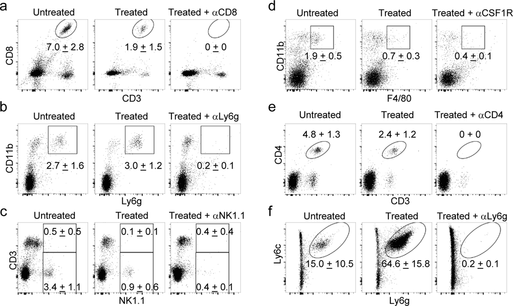
Therapies that synergistically stimulate immunogenic cancer cell death (ICD), inflammation, and immune priming are of great interest for cancer immunotherapy. However, even multi-agent therapies often fail to trigger all of the steps necessary for self-sustaining anti-tumor immunity. Here we describe self-replicating RNAs encapsulated in lipid nanoparticles (LNP-replicons), which combine three key elements: (1) an LNP composition that potently promotes ICD, (2) RNA that stimulates danger sensors in transfected cells, and (3) RNA-encoded IL-12 for modulation of immune cells. Intratumoral administration of LNP-replicons led to high-level expression of IL-12, stimulation of a type I interferon response, and cancer cell ICD, resulting in a highly inflamed tumor microenvironment and priming of systemic anti-tumor immunity. In several mouse models of cancer, a single intratumoral injection of replicon-LNPs eradicated large established tumors, induced protective immune memory, and enabled regression of distal uninjected tumors. LNP-replicons are thus a promising multifunctional single-agent immunotherapeutic.


Figures

Extended Data Fig. 1.
Extended Data Fig. 1.. Comparison of size, zeta potential, RNA encapsulation efficiency, and morphology for the nanoparticles.
a-b, Mean diameter (a) (nm) and Zeta potential (b) (mv) of different nanoparticles with or without encapsulation of replicon RNA (n = 3 /group; error bars are mean ± s.d.). (c) RNA encapsulation efficiency of DOTAP, Lipofectamine, and TT3 nanoparticles (n = 3 /group; error bars are mean ± s.d. n.d. means non-detectabsle. d-g, morphologies of DOTAP and TT3 lipid nanoparticles without (d, f) or with replicon RNA (e, g) were imaged by Cryo-TEM. Shown are representative images from one of two independent experiments. Scale bar (100 nm) is indicated.
Extended Data Fig. 2.
Extended Data Fig. 2.. Immunogenic cell death in human cancer cells and pathways of interferon stimulation in response to treatments with TT3 or TT3-Rep.
a-c, Human A549 lung carcinoma (a), SK-MEL-5 melanoma (b), or Hela cervical cancer cells (c) were treated with 10 μg/mL empty TT3 LNPs (TT3), TT3 LNPs carrying replicons encoding GFP (TT3-Rep), or left untreated as indicated. Three days post treatment, the percentage of viable cells was enumerated (left column). One day post treatment, the ICD markers extracellular ATP (second column), CRT+ cells (third column), extracellular HMGB1 (fourth column) were measured. d, Twelve hours post treatment of 10 μg/mL TT3 LNP (LNP) or TT3 LNP carrying replicons encoding mCherry (LNP-Rep), levels of MDA-5 and TLR3 mRNA in B16F10 cells were assessed by qPCR. Shown are fold changes of MDA-5 and TLR3 expression in different treatments in comparison to untreated cells normalized by actin B expression. e, One day post treatment of A549 type I interferon reporter cells or MDA-5 knockout A549 reporter cells with 10 μg/mL TT3 LNP (LNP) or TT3 LNP carrying replicons encoding mCherry (LNP-mCherry), the supernatants were collected and incubated to detect stimulation the expression of the luciferase reporter. Shown are relative luminescence units. Throughout error bars are mean ± s.d. from a-c (n = 3 /group), d (n = 6 /group), e (n =4 /group); P values were determined by one way (a, b, c) or two way ANOVA (d, e) using PRISM Software and exact P values were indicated.
Extended Data Fig. 3.
Extended Data Fig. 3.. Typical gating strategy for FACS analysis.
C57Bl/6 mice bearing established B16F10 tumors and TDLNs were necropsied and prepared as single cells suspension. The cells were stained with antibodies against cell surface makers as indicated and followed with flow cytometry (see methods). a. Shown are typical gating strategies of granulocytes, M-MDSCs, monocytes, macrophages, CD4 T cells, CD8 T cells, NK cells, NKT cells, mCherry positive cells in tumor cells for Fig. 2d-2g and Fig. 3e. b. Shown are typical gating strategies of CRT positive cells in SSChi FSChi live tumor cells from B16F10 tumor bearing mice treated without or with LNP-Rep for Fig. 2h.
Extended Data Fig. 4.
Extended Data Fig. 4.. Treatments of LNP-Rep encoding mCherry, IL-12-alb, or IL-12-alb-lum reprogrammed tumor and systemic microenvironments with low toxicity.
a, IL12-alb-lum produced by LNP-replicon-transfected tumor cells binds to collagen I. B16F10 cells were transfected with LNP-Rep(IL-12-alb-lum) and the supernatants of the transected cells were added to collagen-coated plates for analysis of binding by ELISA in comparison of standards protein IL12-alb-lum. Shown are the ELISA absorbance for IL-12 detection versus concentration of added IL-12-alb-lum using the supernatants of B16F10 cells that were transfected with LNP-Rep(IL-12-alb-lum) or left untreated (error bars are mean ± s.d. from n = 3 /group). b-c, Comparison of AST (b) and ALT (c) levels in serum at day 1 and 3 after the indicated treatments (error bars are mean ± s.e.m. from n = 5 mice/group). d, Comparison of CXCL9 and CXCL10 levels in tumor at day 1 and 3 after the indicated treatments (error bars are mean ± s.e.m. from n = 5 mice/group). P values were determined by two way ANOVA analysis using PRISM Software and exact P values were indicated.
Extended Data Fig. 5.
Extended Data Fig. 5.. Therapeutic efficacy of LNP-Rep encoding IL-12 in distinct treatment models.
a-b, Groups of C57Bl/6 mice (n=10/group) bearing established B16F10 tumors were treated when tumors reached 50 mm2 with a single injection of 10 μg LNP-rep(IL-12-alb-lum) on day 7 (indicated by red arrows) or with 2.3 μg recombinant IL-12-alb-lum on days 5, 11, and 17 (indicated by blue arrows). Shown are average (mean + s.d.) tumor growth (a) and overall survival (b) over time. c-d, Groups of Balb/c mice bearing established CT26 tumors were treated when tumors reached 50 mm2 with a single injection on day 7 or 3 injections (day 7, 10, 13) of 10 μg LNP-rep(IL-12-alb-lum) start from day 7 (untreated n=6 animals/group, other groups n=8 animals/group). Shown are average (mean + s.e.m.) tumor growth (c) and overall survival (d) over time. e-g, Groups of C57Bl/6 mice bearing established B16F10 tumors were treated when tumors reached 50 mm2 with a single injection of different dosages of the LNP-encapsulated replicon RNA as indicated. Shown are IL-12 levels (mean + s.e.m.) in tumors at one day post injection as measured by ELISA (n=5/group) (e), average (mean + s.d.) tumor growth (f) and overall survival (g) over time with the treatments as indicated (untreated n=6 animals/group, other groups n=8 animals/group). n.d., non-detectable. h-j, C57Bl/6 mice (untreated n=12 mice/group, other groups n=14 mice/group) were inoculated s.c. with 106 and 0.3x10⁶ B16F10 cells in the left and right flanks, respectively. When the left flank tumor reached ~50 mm2 in size, this lesion was injected with 10 μg of the indicated LNP-replicon. Shown is tumor area (mean + s.d.) in each flank (h, i), and animal survival over time (j). P values were determined by one-sided Turkey’s multiple comparison test (Fig. 5f, 5h, and 5i), or by one-sided Log-Rank (Mantel-Cox) test (Fig. 5b, 5d, 5g, and 5j) using PRISM Software and exact P values were indicated.
Extended Data Fig. 6.
Extended Data Fig. 6.. Depletion efficiency in peripheral blood (a-e) and in tumors (f).
C57BL/6J mice bearing B16F10 tumors were administered the indicated depleting antibodies beginning one day prior to injection with 10 μg LNP-Rep(IL12-alb-lum). Then the depletion efficiency in peripheral blood (a-d, Untreated n = 4 mice, treated and untreated n = 5 mice/group; e, n=10 mice/group) and in tumor (f, n=5 mice/group) were determined by flow cytometer. Shown are typical FACS plots from the groups of Untreated, Treated (LNP-Rep(IL-12-alb-lum), and Treated with depleting antibodies as indicated. The mean and standard deviation of gated populations are indicated.
Extended Data Fig. 7.
Extended Data Fig. 7.. CD8 T cells and Batf3 are critical for therapeutic efficacy.
a, The cured mice lost protection while depletion of CD8 T cells. Mice treated as in Fig. 4b that rejected their primary tumor (Naïve n=12 mice/group, Cured (LNP-Rep(IL-12-alb)) n=14 mice/group, Cured (LNP-Rep(IL-12-alb-lum)) n=14 mice/group) were administered antibodies against CD8a beginning one day prior to rechallenge with 0.1x10⁶ B16F10 cells. Then antibody against CD8a was administrated every 3 days and followed for survival mice were observed. b, B16F10 tumor bearing mice in absence of Batf3 decreases responses to treatment of LNP-Rep(IL-12-alb). Shown are tumor areas over time after intratumoral injection of LNP-Rep encoding IL-12-alb into B16F10 tumors (mean + s.e.m.) in C57BL/6J or Batf3−/− mice (B6-Untreated n=5 mice/group, B6-LNP-Rep(IL-12-alb) n=7 mice/group, Baft3−/− -Untreated n=5 mice/group, Baft3−/− -LNP-Rep(IL-12-alb) n=6 mice/group). Throughout, P values were determined by one-sided Log-Rank (Mantel-Cox) test (a), or by one-sided Tukey’s multiple comparison test (b) using PRISM Software and exact P values were indicated.
Extended Data Fig. 8.
Extended Data Fig. 8.. Typical gating strategy for FACS analysis.
C57Bl/6 mice bearing established B16F10 tumors and TDLNs were necropsied and prepared as single cells suspension. The cells were stained with antibodies against cell surface makers as indicated and followed with flow cytometry (see methods). a. Shown are typical gating strategies of conventional DC1 and DC2 in TDLN cell for Fig. 6f. b. Shown are gating strategies of naïve Pmel CD8 T cells activation by cells from inguinal lymph nodes for Fig. 6g.
Figure 1 |
Figure 1 |. Lipid nanoparticle-replicon formulations induce high levels of payload gene expression and immunogenic cell death.
a, Design of lipid nanoparticle/replicon system comprised of a cancer cell-killing lipid formulation encapsulating self-replicating RNA encoding an immunomodulatory gene payload. ICD: immunogenic cell death, TME: tumor microenvironment, TAA: tumor associated antigen. b, Percentages of PI+Annexin V+ B16F10 cells one day post treatment with DOTAP nanoparticles (DOTAP), Lipofectamine (Lipo), TT3 nanoparticles (TT3), Electroporation, DOTAP, Lipo, or TT3 nanoparticles encapsulating replicon RNA (DOTAP-Rep, Lipo-Rep, TT3-Rep) or mutant dead replicon (DOTAP-deRep, Lipo-deRep, and TT3-deRep), or treated with electroporation in the presence of replicon RNA or dead replicon (Electro-Rep and Electro-deRep, respectively). Lipid nanoparticle formulations were added at 10 μg/mL RNA (corresponding to 100 μg/mL TT3 lipids). Shown are mean (box) of different treatments from 3 technical cell culture replicates from one of two independent experiments. c, Viability of B16F10 cells after 3 days incubation with LNP or RNA treatments as indicated (RNA, naked replicon RNA). Shown are mean (box) of different treatments from 3 technical cell culture replicates from one of two independent experiments. d, GFP expression in B16F10 cells at 12-hours post treatment with indicated formulations. Shown are representative flow cytometry plots and mean (box) frequencies of transfected cells from 3 technical cell culture replicates in a single experiment. e-g, B16F10 cells were treated as indicated. Shown are percentages of CRT+ cells (e) and levels of extracellular ATP (f) and HMGB1 (g) at 1 day post transfection. Shown are mean (box) of different treatments from 3 technical cell culture replicates from one of two independent experiments. h-i, HEK-blue TLR reporter and RAW-Lucia-ISG cells were transfected with nanoparticles with or without encapsulated replicon RNA (10 μg/mL RNA). TLR2, 3, 4, 7 and 9 activation as indicated by secreted alkaline phosphatase reporter was assessed by absorbance (h) and secretion of luciferase for Raw-Lucia-ISG was determined by luminescence (i). Shown are mean (box) of different treatments from 3 technical cell culture replicates from one of two independent experiments. j, Quantitative analysis of Stat1, Stat2, IRF9, IRF3, and cGAS mRNA transcripts by qPCR in B16F10 cells at 12 hours post transfection with formulations as indicated. Shown are mean (box) of different treatments from 3 technical cell culture replicates from one of two independent experiments. The source data of Fig. 1a-1j are found in Li_SourceData_Fig.1.
Figure 2 |
Figure 2 |. TT3 lipid nanoparticles carrying replicon RNA induce immunogenic cell death and immune infiltration of tumors in vivo.
a-b, Groups of C57Bl/6 mice were inoculated with 106 B16F10 cancer cells in the flank and treated when tumors reached 50 mm2 with a single injection of empty LNP (n=8 mice/group), 10 μg LNP-Rep (n=9 mice/group), 10 μg LNP-deRep (n=9 mice/group), or left untreated (n = 8 mice/group). Shown are average tumor growth (mean ± s.d.) (a) and overall survival (b) over time. c-d, Enumeration (mean ± s.e.m.) of tumor-infiltrating granulocytes (c) and other leukocyte populations (d) 3 days after injection of various formulations as indicated (n = 5 mice/group). e, Uptake (mean ± s.e.m. from n = 5 mice/group) of Cy5-labeled TT3 LNP-Rep by tumor-infiltrating lymphocytes (TILs) and cancer cells assessed by flow cytometry 12 hr post injection. f, Expression (mean ± s.e.m. from n = 5 mice/group) of mCherry 12 hours post injection in CD45- cancer cells and TILs. g, Cell viability (mean ± s.e.m. from n = 5 mice/group) in tumors 3 days after TT3 LNP-Rep injection. h, Percentages (mean ± s.e.m. from n = 5 mice/group) of CRT+ cells in tumors at 1 and 3 days post injection as indicated. i, IFN-α2 expression (mean ± s.e.m. from n = 5 mice/group) in tumors at day 1 and day 3 post injection of LNP-Rep as measured by ELISA. j, qPCR analysis (mean ± s.d. from n = 8 (4 mice/group and two technical replicates/mouse) of Stat1, Stat2, IRF9, IRF3, and cGAS mRNA transcripts in tumors at 1 day post LNP-Rep injection. P values were determined by one-sided Tukey’s multiple comparison test (Fig. 2a), or by Log-Rank (Mantel-Cox) test (Fig. 2b), or by one-way (Fig. 2c) or two-way (Fig. 2d, 2e, 2f, 2h, 2i and 2j) ANOVA analysis, or by two-sided Student’s T-test (2g) using GraphPad PRISM Software and exact P values were indicated. The source data of Fig. 2a-2j are found in Li_SourceData_Fig. 2.
Figure 3 |
Figure 3 |. LNP-replicons encoding IL-12-alb or IL-12-alb-lum remodel the tumor microenvironment.
a, 0.5×10⁶ B16F10 cells were transfected with LNP-Rep encoding mCherry, IL-12-alb, or IL-12-alb-lum and cultured in 1 ml media. Shown are concentrations (mean ± s.e.m.) of IL-12 (ng/ml) in the supernatants of 3 technical cell culture replicates at 24 hr from one of two independent experiments. b-g, C57Bl/6 mice (n = 5 mice/group) were inoculated with B16F10 tumors and treated with LNP-Rep encoding mCherry, IL-12-alb, or IL-12-alb-lum when tumors were ~50 mm2 as in Fig. 2. b, IL-12 levels (mean ± s.e.m.) in tumors were assessed at 1, 3, or 7 days post replicon injection. c, Cytokines and chemokine in serum at 1 and 3 days post injection with LNP-Reps encoding mCherry, IL-12-alb, or IL-12-alb-lum as measured by Luminex ELISA. d, Changes in body weight (mean ± s.e.m.) over time post tumor inoculation. e, Immune composition (mean ± s.e.m.) of leukocytes in tumors at day 3 post replicon injection. f, Luminex analysis of protein expression in tumors at 1 and 3 days post replicon injection. Shown are clustered protein levels as a heat map. g, Representative images of tumor sections at 3 days post replicon injection as indicated from one of two independent experiments. Shown is staining for nuclei (DAPI, blue) and CD31 (green). Red boxes at left highlight regions in the interior and border of the tumors shown in the high magnification images at right. Scale bars used in global and zoom in images are 1000 and 100 μm as indicated. P values were determined one way (Fig. 3a) or two way (Fig. 3b, 3e) ANOVA analysis or one-sided Tukey’s multiple comparison test (Fig. 3d) using GraphPad PRISM Software and exact P values were indicated. The source data of Fig. 3a-3f are found in Li_SourceData_Fig. 3. The source data of Fig. 3g are stored in Figshare.
Figure 4 |
Figure 4 |. A single injection of LNP-replicons encoding IL-12-alb or IL-12-alb-lum can eradicate large established tumors
a-f, Tumor area (mean ± s.e.m.) (a, c, e) and mouse survival (b, d, f) over time after one intratumoral injection of 10 μg LNP-replicon into ~50 mm2 B16F10 (a-b, Untreated n=10 mice/group, LNP-Rep n=9 mice/group, LNP-Rep(IL-12-alb) n=12 mice/group, LNP-Rep(IL-12-alb-lum) n=11 mice/group), YUMMER1.7 (c-d, Untreated n=7 mice/group, LNP-Rep(IL-12-alb-lum) n=8 mice/group), or CT26 tumors (e-f, Untreated n=7 mice/group, LNP-Rep n=7 mice/group, LNP-Rep(IL-12-alb) n=8 mice/group, LNP-Rep(IL-12-alb-lum) n=8 mice/group), respectively. P values were determined by one-sided Tukey’s multiple comparison test (Fig. 4a, 4e), or by one-sided Log-Rank (Mantel-Cox) test (Fig. 4b, 4d, and 4f) using GraphPad PRISM Software and exact P values were indicated. The source data of Fig. 4a-4f are found in Li_SourceData_Fig. 4.
Figure 5 |
Figure 5 |. Local LNP-replicon therapy regresses distal untreated tumors and eliminates metastases.
a-d, C57Bl/6 mice (Untreated n=7 mice/group, Untreated + α-PD1 n=8 mice/group, LNP-Rep (IL-12-alb) n=8 mice/group, LNP-Rep (IL-12-alb-lum) + α-PD1 n=8 mice/group) were inoculated s.c. with 106 and 0.3×10⁶ B16F10 cells in the left and right flanks, respectively. When the left flank tumor reached ~50 mm2 in size, this lesion was injected with 10 μg of the indicated LNP-replicon and systemic anti-PD-1 antibody was initiated, dosed every 3 days. Shown is the experimental setup (g), tumor area (mean ± s.d.) in each flank (h, i), and animal survival over time (j). e-h, C57Bl/6 mice (Untreated (s.c.) n= 12 mice/group, Untreated (s.c. + i.v.) n= 12 mice/group, LNP-Rep(IL-12-alb) (s.c.) n= 10 mice/group, LNP-Rep(IL-12-alb-lum) (s.c.) n= 10 mice/group) were injected with 1 million B16F10 cells s.c. in the flank. Two days later, an additional 0.25 million B16F10 cells were injected i.v. On day 7, flank tumors were injected with 10 μg LNP-Rep(IL-12-alb) or LNP-Rep(IL-12-alb-lum) as indicated. Shown is the experimental setup (e), tumor area (mean ± s.d.) of the treated flank tumor (f), enumeration of tumor nodules (mean ± s.e.m.) in the lungs at the indicated times (g), and representative images of lungs at indicated time points (h). Scale bars (1 cm) are as indicated. P values were determined by one-sided Tukey’s multiple comparison test (Fig. 5b, 5c, and 5f), or by one-sided Log-Rank (Mantel-Cox) test (Fig. 5d), or by one way ANOVA analysis (Fig. 5g) using GraphPad PRISM Software and exact P values were indicated. The source data of Fig. 5b-5d, 5f-5g are found in Li_SourceData_Fig. 5. Source data of Fig. 5h are found in SourceData_Li_Fig.5h-Lungimages.pdf and also stored in Figshare.
Figure 6 |
Figure 6 |. Cellular and molecular pathways governing LNP-replicon therapeutic efficacy.
a-d, C57Bl/6 mice bearing B16F10 tumors (a-b, Untreated n=9 mice/group, Treated n=10 mice/group, Treated + α-NK1.1 n=9 mice/group, Treated + α-Ly6g n=10 mice/group, Treated + α-CSF1R n=10 mice/group; c-d, n=10 mice/group) were administered the indicated depleting antibodies beginning one day prior to injection with 10 μg LNP-Rep(IL-12-alb-lum) and every 3 days thereafter. Shown are tumor areas (mean ± s.e.m.) (a, c) and mouse survival over time (b, d). e, Mice treated as in Fig. 3h that rejected their primary tumor (n=9 mice/group) were rechallenged with 0.1×10⁶ B16F10 cells, and observed for survival. f, Mice bearing B16F10 tumors (n=5 mice/group) were treated as in Fig. 3h, and cDC1 (mean ± s.e.m.) were enumerated in TDLNs 1 day after replicon injection. g, Mice bearing B16F10 tumors (n=4 mice/group) were treated as in Fig. 3h, and 3 days post replicon injection, TDLN cells were recovered and co-cultured with Pmel reporter CD8 T cells. Shown is CD69 (mean ± s.e.m.) upregulation on responding pmel CD8 T cells. h-j, B16F10 cancer cells were inoculated in WT, Batf3−/−, Myd88−/−, or STING−/− mice (h, B6-Untreated n=5 mice/group, B6-LNP-Rep(IL-12-alb) n=7 mice/group, Batf3−/−-Untreated n=5 mice/group, Batf3−/−- LNP-Rep(IL-12-alb) n=6 mice/group; i-j, B6-Untreated n=7 mice/group, B6-LNP-Rep(IL-12-alb) n=8 mice/group, Myd88−/−-Untreated n=6 mice/group, Myd88−/−-LNP-Rep(IL-12-alb) n=6 mice/group, STING−/−-Untreated n=8 mice/group, STING−/−-LNP-Rep(IL-12-alb) n=9 mice/group), and treated when tumors reached 50 mm2 with a single injection of the indicated LNP-replicons. Shown is tumor area (mean ± s.e.m.) or animal survival over time. P values were determined by one-sided Tukey’s multiple comparison test (Fig. 6a, 6c, and 6i), or by one-sided Log-Rank (Mantel-Cox) test (Fig. 6b, 6d, 6e, 6h, and 6j), or by one way ANOVA analysis (Fig. 6f and 6g) using GraphPad PRISM Software and exact P values were indicated. Source data of Fig. 6a-6j are found in Li_SourceData_Fig. 6.

Comment in

  • Pseudovirus for immunotherapy.
    Kepp O, Kroemer G. Kepp O, et al. Nat Cancer. 2020 Sep;1(9):860-861. doi: 10.1038/s43018-020-00107-2. Nat Cancer. 2020. PMID: 35121951 No abstract available.

References

    1. Sharma P & Allison JP The future of immune checkpoint therapy. Science 348, 56–61, doi:10.1126/science.aaa8172 (2015). - DOI - PubMed
    1. Zou W, Wolchok JD & Chen L PD-L1 (B7-H1) and PD-1 pathway blockade for cancer therapy: Mechanisms, response biomarkers, and combinations. Sci Transl Med 8, 328rv324, doi:10.1126/scitranslmed.aad7118 (2016). - DOI - PMC - PubMed
    1. Wolchok JD et al.Overall Survival with Combined Nivolumab and Ipilimumab in Advanced Melanoma. N Engl J Med 377, 1345–1356, doi:10.1056/NEJMoa1709684 (2017). - DOI - PMC - PubMed
    1. Chen DS & Mellman I Elements of cancer immunity and the cancer-immune set point. Nature 541, 321–330, doi:10.1038/nature21349 (2017). - DOI - PubMed
    1. Hammerich L et al.Systemic clinical tumor regressions and potentiation of PD1 blockade with in situ vaccination. Nat Med 25, 814–824, doi:10.1038/s41591-019-0410-x (2019). - DOI - PubMed

Publication types