. 2021 Sep 1;25(5):459-466.
doi: 10.4196/kjpp.2021.25.5.459.

Dronedarone hydrochloride enhances the bioactivity of endothelial progenitor cells via regulation of the AKT signaling pathway

Affiliations

Dronedarone hydrochloride enhances the bioactivity of endothelial progenitor cells via regulation of the AKT signaling pathway

Jian Zhang et al. Korean J Physiol Pharmacol. .

Abstract

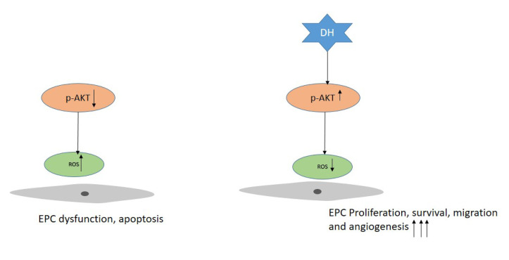
Cardiovascular disease (CVD) and its complications are the leading cause of morbidity and mortality in the world. Because of the side effects and incomplete recovery from current therapy, stem cell therapy emerges as a potential therapy for CVD treatment, and endothelial progenitor cell (EPC) is one of the key stem cells used for therapeutic applications. The effect of this therapy required the expansion of EPC function. To enhance the EPC activation, proliferation, and angiogenesis using dronedarone hydrochloride (DH) is the purpose of this study. DH received approval for atrial fibrillation treatment and its cardiovascular protective effects were already reported. In this study, DH significantly increased EPC proliferation, tube formation, migration, and maintained EPCs surface marker expression. In addition, DH treatment up-regulated the phosphorylation of AKT and reduced the reactive oxygen species production. In summary, the cell priming by DH considerably improved the functional activity of EPCs, and the use of which might be a novel strategy for CVD treatment.

Keywords: Akt; Cardiovascular disease; Dronedarone; Endothelial progenitor cells.


Conflict of interest statement

CONFLICTS OF INTEREST

The authors declare no conflicts of interest.

Figures

Fig. 1
Fig. 1. Effect of DH on EPC proliferation.
(A) The chemical structure of DH. (B) EPCs were cultured with varying concentrations of DH for 24 h, 48 h and cell proliferation assessed by the CCK-8 assay. (C) EPC morphology during treatment with DH. Data represent the mean ± SEM. DH, dronedarone hydrochloride; EPC, endothelial progenitor cell. The results are considered statistically significant at *p < 0.05, **p < 0.01, ***p < 0.001 when compared to untreated groups.
Fig. 2
Fig. 2. Characterization of endothelial functional activation by EPC and EC surface markers.
The expression of EPC (CD34, c-kit, CXCR4) and EC (Tie2, CD31, CD144) surface markers were analyzed by flow cytometry, and no significant differences were observed. FACS gating was applied using unstained cells as a negative control. The fraction of positively stained cells was determined by comparison with unstained cells. The percentage of positively stained cells is indicated by the positive peaks (red lines show cells stained with each antibody, and black lines show the negative control cells). EPC, endothelial progenitor cell; EC, endothelial cell; FACS, fluorescence-activated cell sorting.
Fig. 3
Fig. 3. DH enhances EPC functional ability.
(A) EPCs treated with or without DH (0.5 µM) were seeded on matrigel-coated wells and the angiogenic function was evaluated in a tube formation assay. Representative images of tube formation (40×). (B, C) Quantification of the number of branches and length. (D, E) Representative images of the scratch wound healing and migration assay. (F, G) Quantification of the migrated area and number of migrated cells. Data represent the mean ± SEM. DH, dronedarone hydrochloride; EPC, endothelial progenitor cell. The results are considered statistically significant at *p < 0.05, **p < 0.01, ***p < 0.001 when compared to untreated groups.
Fig. 4
Fig. 4. DH enhances angiogenic activity by regulating AKT signaling.
(A) Protein level expression of AKT and its phosphorylation was evaluated by Western blotting in dose dependent-manner of DH. (B) Quantification of Western blots. (C) Protein level expression of AKT and its phosphorylation was evaluated by Western blots. EPCs were cultured with DMSO, DH, and DH plus AKT inhibitor (5 µM). (D) Quantification of Western blots. (E) Cell treated with DH (0.5 µM) and in the combination with AKT inhibitor (5 µM) for 24 h then proliferation was measured. Scratch wound healing assay was performed to measure wound healing properties and cell migration was measured with the Image J software (NIH, Bethesda, MD, USA). (F) Representative images of the scratch wound healing assay. (H) Carboxy-H2DFFDA was used to measure ROS production. Cells were pretreated with DH with or without AKT inhibitor then ROS was measured by FACS. (I) Quantification of ROS production. Data represent the mean ± SEM. DH, dronedarone hydrochloride; EPC, endothelial progenitor cell; ROS, reactive oxygen species; FACS, fluorescence-activated cell sorting. The results are considered statistically significant at *p < 0.05, **p < 0.01, ***p < 0.001 when compared to untreated groups.
Fig. 5
Fig. 5. Schematic diagram representation of DH enhance the angiogenic activity via AKT signaling.
Exposure of DH up-regulate the expression of p-AKT, ROS reduction lead to the improvements of EPC function. DH, dronedarone hydrochloride; EPC, endothelial progenitor cell; ROS, reactive oxygen species.

References

    1. Cully M. Cardiovascular disease: BET inhibitor attenuates heart failure. Nat Rev Drug Discov. 2017;16:453. doi: 10.1038/nrd.2017.125. - DOI - PubMed
    1. Dimmeler S. Cardiovascular disease review series. EMBO Mol Med. 2011;3:697. doi: 10.1002/emmm.201100182. - DOI - PMC - PubMed
    1. Kuehn BM. Sleep duration linked to cardiovascular disease. Circulation. 2019;139:2483–2484. doi: 10.1161/CIRCULATIONAHA.119.041278. - DOI - PubMed
    1. Yoshida Y, Yamanaka S. Recent stem cell advances: induced pluripotent stem cells for disease modeling and stem cell-based regeneration. Circulation. 2010;122:80–87. doi: 10.1161/CIRCULATIONAHA.109.881433. - DOI - PubMed
    1. Asahara T, Murohara T, Sullivan A, Silver M, van der Zee R, Li T, Witzenbichler B, Schatteman G, Isner JM. Isolation of putative progenitor endothelial cells for angiogenesis. Science. 1997;275:964–967. doi: 10.1126/science.275.5302.964. - DOI - PubMed