Review
. 2022 Jan;43(1):225-232.
doi: 10.1007/s00246-021-02710-1. Epub 2021 Aug 27.

Topsy-Turvy Heart with Aortopulmonary Window and Severe Airway Malacia: Prenatal Diagnosis and Review of the Literature

Affiliations
Review

Topsy-Turvy Heart with Aortopulmonary Window and Severe Airway Malacia: Prenatal Diagnosis and Review of the Literature

Yahia Hejazi et al. Pediatr Cardiol. 2022 Jan.

Abstract

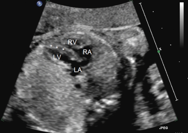
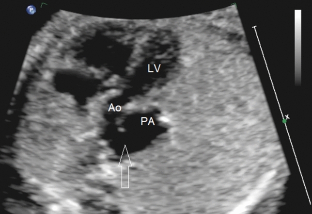
The topsy-turvy heart is a very rare cardiac malformation that involves a global 90° clockwise rotation of the heart along its long axis. This rotation results in the displacement of the great arteries and severe elongation and stretching of the brachiocephalic arteries and the bronchi. We present an unusual case of topsy-turvy heart diagnosed prenatally with a large aorto-pulmonary window and. This case gives an insight into the morphological details and clinical presentation of this rare malformation and its associated complications. We also present a review of the literature of this rare anomaly showing only 15 live cases that have been published with only three cases diagnosed prenatally.

Keywords: Aortopulmonary window; Superior–inferior ventricle; Topsy-turvy heart; Vascular anomalies.


Conflict of interest statement

The authors declare that they do not have any potential conflicts of interest.

Figures

Fig. 1
Fig. 1
Fetal echocardiographic four-chamber view showing the abnormal horizontal position of the interventricular septum (*) as a result of the superior inferior rotation of the heart. LV left ventricle, RV right ventricle, LA left atrium, RA right atrium
Fig. 2
Fig. 2
Fetal long axis echocardiographic view with 2D and color showing the superior–inferior relationship of the ventricles, the short ascending aorta, and an elongated subclavian artery. LV left ventricle, RV right ventricle, Ao aorta, SCA subclavian artery
Fig. 3
Fig. 3
Fetal echocardiographic view showing the left outflow tract and a large AP window (arrow) connecting the aorta with the pulmonary artery. Ao aorta, PA pulmonary artery, LV left ventricle
Fig. 4
Fig. 4
MDCTA reconstructed images. A Lateral 3DVR demonstrating superior RV and inferior LV with an APW. B Posterior 3DVR reveals four elongated head and neck arterial branches from the posteroinferior aortic arch and abrupt high termination of the abdominal descending aorta. C Coronal MinIP (lung window) reconstruction showing ribbon-like stretched right bronchus intermedius (arrowhead), collapsed proximal left main bronchus with bilateral upper lobe atelectasis (*), and ball-valve type hyperinflation of the remainder of the lungs. MDCTA multidetector CT angiogram, 3DVR three-dimensional volume rendering, APW aortopulmonary window, LCCA left common carotid artery, RCCA right common carotid artery, LSCA left subclavian artery, RSCA right subclavian artery, MinIP minimum intensity projection, superior RV and inferior LV superinfero ventricles
Fig. 5
Fig. 5
A Aortic angiogram with anteroposterior and lateral views: showing dextroposition, cardiac apex abnormally oriented to left upper thorax. Catheter course as following: inferior vena cava, right atrium, right ventricle, pulmonary artery, then through AP window to the aorta. Aortogram showing significant elongation of the head and neck vessels with abnormal take-off at level of diaphragm (contrast is filling only right common carotid artery in this image). B Aortogram, anteroposterior view, showing interruption of abdominal aorta, and multiple abnormal collateral vessels supplying the abdominal organs

References

    1. Freedom RM, Culham J, Moes C. Superoinferior ventricles: a consideration of so-called criss-cross atrioventricular connections. In: Freedom RM, Culham J, Moes C, editors. Angiography of congenital heart disease. New York: Macmillan Publishing Company; 1984. pp. 629–642.
    1. Freedom RM, Mawson JB, Yoo SJ, Benson LN. Twisted atrioventricular connections. So-called superoinferior ventricles or criss-cross heart. In: Freedom RM, Mawson JB, Yoo SJ, Benson LN, editors. Congenital heart disease: textbook of angiography. Armonk: Futura Publishing Company; 1997. pp. 1313–1333.
    1. Anderson RH, Yoo SJ. Abnormal positions and relationship of the heart. In: Anderson RH, Baker EJ, Penny D, Redington AN, Rigby ML, editors. Pediatric cardiology. 3. Philadelphia: Churchill Livingstone: Elsevier; 2010. pp. 991–1001.
    1. Sasikumar D, Dharan BS, Menon S, Sivasubramanian S, Kapilamoorthy TR. Surgical closure of aortopulmonary window in a topsy-turvy heart: a surgical challenge. Ann Thorac Surg. 2016;101(5):1982–1984. doi: 10.1016/j.athoracsur.2015.04.128. - DOI - PubMed
    1. Alkhateeb A, Mansour A, Tanidir IC. Topsy-turvy heart: a first recorded case report with intracardiac anomaly. Echocardiography. 2021;38(1):114–117. doi: 10.1111/echo.14950. - DOI - PubMed