. 2021 Dec;89(12):1633-1646.
doi: 10.1002/prot.26223. Epub 2021 Sep 6.

Computational models in the service of X-ray and cryo-electron microscopy structure determination

Affiliations

Computational models in the service of X-ray and cryo-electron microscopy structure determination

Andriy Kryshtafovych et al. Proteins. 2021 Dec.

Abstract

Critical assessment of structure prediction (CASP) conducts community experiments to determine the state of the art in computing protein structure from amino acid sequence. The process relies on the experimental community providing information about not yet public or about to be solved structures, for use as targets. For some targets, the experimental structure is not solved in time for use in CASP. Calculated structure accuracy improved dramatically in this round, implying that models should now be much more useful for resolving many sorts of experimental difficulties. To test this, selected models for seven unsolved targets were provided to the experimental groups. These models were from the AlphaFold2 group, who overall submitted the most accurate predictions in CASP14. Four targets were solved with the aid of the models, and, additionally, the structure of an already solved target was improved. An a posteriori analysis showed that, in some cases, models from other groups would also be effective. This paper provides accounts of the successful application of models to structure determination, including molecular replacement for X-ray crystallography, backbone tracing and sequence positioning in a cryo-electron microscopy structure, and correction of local features. The results suggest that, in future, there will be greatly increased synergy between computational and experimental approaches to structure determination.

Keywords: CASP; X-ray crystallography; cryo-EM; protein structure prediction.


Figures

Figure 1.
Figure 1.
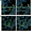
(A) Partial FoxB model obtained by experimental phasing before the CASP14 model became available. At this point the model could not be further improved and the project was stuck for a year. (B) Experimental phases with partial FoxB model (map shown at 1.2σ level).
Figure 2.
Figure 2.
Workflow of FoxB structure determination. The structure was determined by MR-SAD using the AlphaFold2 model and experimental phases. (A) Anomalous difference map with Se and Fe sites at 2σ. (B) Overall map of FoxB after refinement (2σ). (C) Superposition of the final model (green) and AlphaFold2 model (cyan) shows excellent agreement. Density for heme groups (not present in AlphaFold2 model) is shown.
Figure 3.
Figure 3.
AlphaFold2 models of AR9 nvRNAP proteins fit the cryo-EM density nearly perfectly. The cryo-EM-derived structures of gp105, gp154, and two gp226 domains are colored according to the color code given in the upper left corner of each panel. All AlphaFold2 models are colored magenta. The electron density is contoured at 4.25 standard deviations above the mean and colored semi-transparent grey. Regions where no cryo-EM-derived structure existed prior to the availability of the AlphaFold2 models are indicated with a dashed line and their boundary residues are labeled.
Figure 4.
Figure 4.
Inaccuracies in AlphaFold2 models. Cryo-EM-derived structures and AlphaFold2 models of several AR9 nvRNAP subunits are superimposed and regions where the conformation of the AlphaFold2 model deviates significantly from the cryo-EM-derived structure are indicated with a dashed line and their boundary residues are labeled. Note that the folds of both the N- and C-terminal domains of gp226 were predicted correctly, but the structure of the interdomain linker and the relative orientation of the two domains were incorrect.
Figure 5.
Figure 5.
(A) The structure of TSP4-N homo-trimer with each subunit in different color. The dash lines indicate structurally disordered linkers between XD2 and XD3. (B) Superposition of XD2 as seen in the crystal structure (magenta) and the structure predicted by group 427 (green) and group 226 (sky blue). (C) Superposition of AD crystal structure (magenta) and the structure predicted by group 427 (green).
Figure 6.
Figure 6.
Polypeptide chain tracing errors that were corrected by examination of the AlphaFold2 (group 427) structure. (A) The incorrect model in the vicinity of two neighboring proline residues (Pro236 and Pro239) together with the associated difference electron density map with the coefficient 2Fo-Fc colored blue (left) and the model corrected based on the AlphaFold2 predicted structure with the associated 2Fo-Fc difference electron density map (right). The cis bond conformations are highlighted in green (B) The incorrect placement of Ile247 with the associated 2Fo-Fc difference electron density map colored blue and the Fo-Fc difference electron density map colored green (left). Correcting the positions of Pro236 and Pro239 allowed placement of Tyr249 instead of Ile247 and eliminated the residual Fo-Fc difference electron density (right).
Figure 7.
Figure 7.
The crystal structure of dimeric Af1503 (grey) is shown in a superposition with the best AlphaFold2 model (green, monomer). The only noteworthy difference between the prediction and the crystal structure is found in a loop in the PAS domain, which was found to coordinate an ion in the crystal structure.
Figure 8.
Figure 8.
Superposition of Sia24 predictive and crystallographic models. The structure of Sia24 (dark blue) was solved with initial phase determination by molecular replacement using a model generated by AlphaFold2 (yellow).

References

    1. Schwede T, Sali A, Honig B, et al. Outcome of a workshop on applications of protein models in biomedical research. Structure. 2009;17(2):151–159. - PMC - PubMed
    1. Tramontano A The role of molecular modelling in biomedical research. FEBS Lett. 2006;580(12):2928–2934. - PubMed
    1. Kuhlman B, Bradley P. Advances in protein structure prediction and design. Nat Rev Mol Cell Biol. 2019;20(11):681–697. - PMC - PubMed
    1. Simpkin AJ, Thomas JMH, Simkovic F, Keegan RM, Rigden DJ. Molecular replacement using structure predictions from databases. Acta Crystallogr D Struct Biol. 2019;75(Pt 12):1051–1062. - PMC - PubMed
    1. Case DA. Using quantum chemistry to estimate chemical shifts in biomolecules. Biophys Chem. 2020;267:106476. - PMC - PubMed

Publication types