. 2021 Aug 27;15(8):e0009728.
doi: 10.1371/journal.pntd.0009728. eCollection 2021 Aug.

Infecting mosquitoes alters DENV-2 characteristics and enhances hemorrhage-induction potential in Stat1-/- mice

Affiliations

Infecting mosquitoes alters DENV-2 characteristics and enhances hemorrhage-induction potential in Stat1-/- mice

Ka Wan Cheang et al. PLoS Negl Trop Dis. .

Abstract

Dengue is one of the most prevalent arthropod-borne viral diseases in humans. There is still no effective vaccine or treatment to date. Previous studies showed that mosquito-derived factors present in saliva or salivary gland extract (SGE) contribute to the pathogenesis of dengue. In this study, we aimed to investigate the interplay between mosquito vector and DENV and to address the question of whether the mosquito vector alters the virus that leads to consequential disease manifestations in the mammalian host. DENV2 cultured in C6/36 cell line (culture-DENV2) was injected to Aedes aegypti intrathoracically. Saliva was collected from infected mosquitoes 7 days later. Exploiting the sensitivity of Stat1-/- mice to low dose of DENV2 delivered intradermally, we showed that DENV2 collected in infected mosquito saliva (msq-DENV2) induced more severe hemorrhage in mice than their culture counterpart. Msq-DENV2 was characterized by smaller particle size, larger plaque size and more rapid growth in mosquito as well as mammalian cell lines compared to culture-DENV2. In addition, msq-DENV2 was more efficient than culture-DENV2 in inducing Tnf mRNA production by mouse macrophage. Together, our results point to the possibility that the mosquito vector provides an environment that alters DENV2 by changing its growth characteristics as well as its potential to cause disease.


Conflict of interest statement

The authors have declared that no competing interests exist.

Figures

Fig 1
Fig 1. Scheme depicting study design.
The experiment included four groups. (A, B) Inocula prepared in (A) were given to 4 groups of mice as in (B). (A) DENV2-16681 was propagated in C6/36 insect cell line and culture supernatants were collected and virus titer was determined by plague assay (culture-DENV2). Culture-DENV2 was mixed with saliva collected from naïve Aedes aegypti (culture-DENV + naïve saliva). A. aegypti was intrathoracically inoculated with 69 nl of DENV-2 16681 (6.9 × 10² PFU in 69 nl) and saliva was collected on day 7 after infection (infectious saliva). Culture-DENV2 was mixed with infectious saliva that was exposed to ultraviolet (UV) light (culture-DENV2 + UV-inactivated infectious saliva). The titer of DENV2 from either cell culture or infectious mosquitoes was determined by plaque assay and adjusted to 6 × 10³ PFU in 400 μl. (B) Four groups of Stat1-/- mice were injected intradermally at 4 different sites on the upper back with inoculum prepared in (A) in a total volume of 400 μl. Hemorrhage development was observed on day 6 after inoculation.
Fig 2
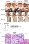
Fig 2. DENV2 from infectious mosquito saliva is more efficient in inducing hemorrhage than DENV2 grown in mosquito cell culture.
Four groups of mice were inoculated with DENV2 following the protocol as described in Fig 1. (A) Mouse dorsal skin, subcutaneous tissues, and abdominal skin were dissected on day 6 after inoculation. The abdominal skin was magnified. (B) Hemorrhaged areas in the abdominal skin were quantified by ImageJ. Percentage of hemorrhaged area was obtained by dividing the sum of the hemorrhaged area in the selected region by the total selected area of the region. Approximately 60% of the total hemorrhaged areas of the abdominal skin was measured. Data presented in bar graphs are the mean ± SD obtained from four independent experiments (mice injected with culture-DENV2, n = 5; mice injected with culture-DENV2 + naïve saliva, n = 11; mice injected with infectious saliva, n = 6; mice injected with culture-DENV2 + UV-inactivated infectious saliva, n = 8). *p < 0.05, **p < 0.01, ***p < 0.005, as analyzed by one-way ANOVA with Dunn’s multiple comparisons test, comparing mice injected with infectious saliva with those injected with culture-DENV2, culture-DENV2 mixed with naïve saliva and culture-DENV2 mixed with UV-inactivated infectious saliva. (C) Dorsal and abdominal skins were fixed in formaldehyde, cut into 4 μm sections and stained with hematoxylin-eosin (H&E) stain. Filled triangles point to extravascular RBC and empty triangles to mononuclear cells. Area in the square was magnified showing red blood cell extravasation and leukocyte infiltration.
Fig 3
Fig 3. DENV2 isolated from infectious mosquito saliva retains hemorrhage-induction potential after one passage in mosquito cell line.
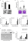
Msq-DENV2 was propagated in C6/36 cell line. Culture supernatants containing the virus (msq-DENV-P1) was titered. Stat1-/- mice were injected intradermally with 6 × 10³ PFU in 400 μl as described above. (A) Dorsal skin, subcutaneous tissues, and abdominal skin were observed on day 6 after inoculation. Representative photos of one mouse from each group is shown. Area in the square in abdominal skin was magnified. (B) Hemorrhaged areas were quantified by ImageJ. Percentage of hemorrhaged area was calculated as in Fig 2. Approximately 60% of the total hemorrhaged areas of the abdominal skin was measured. Data presented in the bar graphs are the mean ± SD obtained from 3 mice. ***p < 0.005, as analyzed by Wilcoxon–Mann–Whitney test, comparing mice inoculated with culture-DENV2 and msq-DENV2-P1.
Fig 4
Fig 4. DENV2 isolated from infectious mosquito saliva are smaller in size, forms larger plaques and grows faster than DENV2 grown in mosquito cell culture.
(A) Dengue virions from culture-DENV2 and msq-DENV2-P1 were observed by transmission electron microscopy. The diameter of dengue virions was measured by ImageJ. Representative images are shown below the bar graph and S2 Fig. n = 14 for each group. *** p < 0.005, by comparing culture-DENV2 with msq-DENV2-P1, was analyzed by Wilcoxon–Mann–Whitney test. Arrowheads point to virions. (B) Culture-DENV2 and infectious saliva were added onto BHK-21 cell monolayers separately. Five days later, plaque size was measured by ImageJ. Data presented in the bar graph are the mean ± SD obtained from 3 independent experiments and analyzed by Wilcoxon–Mann–Whitney test. Representative images are shown below the graph and in S3 Fig. (C) BHK-21 cells were infected with culture-DENV2 or msq-DENV2-P1 at a MOI of 0.01. C6/36 and ATC10 cells were infected with culture-DENV2 or msq-DENV2-P1 at a MOI of 0.1. Viral titers were determined by plaque assay. One-way ANOVA with Dunn’s multiple comparisons test was used to compare the differences in viral titers between culture-DENV2 and msq-DENV2-P1 with data from multiple time points. (D) ATC10 culture supernatants collected at different time points were analyzed for E protein expression by Western blot (upper gel). Coomassie blue staining shows the total protein loaded onto each lane (bottom gel). The relative intensity of E protein in the supernatants is presented in the bar graphs as mean ± SD pooled from 4 independent experiments. n.d. = non-detected, *p < 0.05, **p < 0.01, ***p < 0.005 comparing culture-DENV2 with msq-DENV2-P1. One-way ANOVA with Dunn’s multiple comparisons test was used to compare the differences in E protein intensity between culture-DENV2 and msq-DENV2-P1 with data from multiple time points.
Fig 5
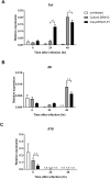
Fig 5. Comparing msq-DENV2 and culture-DENV2 for their ability to induce cytokines in mouse macrophages.
Peritoneal macrophages were obtained from Stat1-/- mice on day 4 after intraperitoneal injection of thioglycollate (Thio-pMac). Thio-pMac were stimulated with culture-DENV2 or msq-DENV2-P1 at a MOI of 20 for 6, 24 or 48 h. RNA was extracted, reverse-transcribed to cDNA for real-time PCR quantitation. mRNA of each cytokine was normalized against mouse Gapdh. n = 4 for each group for each time point. n.s. = non-significant, ***p < 0.005, as analyzed by one-way ANOVA with Dunn’s multiple comparisons test comparing cytokine levels induced by culture-DENV2 and msq-DENV2-P1 from different time points.

References

    1. Bhatt S, Gething PW, Brady OJ, Messina JP, Farlow AW, Moyes CL, et al.. The global distribution and burden of dengue. Nature. 2013;496(7446):504–7. doi: 10.1038/nature12060 - DOI - PMC - PubMed
    1. Cucunawangsih Lugito NPH. Trends of Dengue Disease Epidemiology. Virology: research and treatment. 2017;8:1178122x17695836. - PMC - PubMed
    1. Pinheiro FP, Corber SJ. Global situation of dengue and dengue haemorrhagic fever, and its emergence in the Americas. World health statistics quarterly Rapport trimestriel de statistiques sanitaires mondiales. 1997;50(3–4):161–9. - PubMed
    1. Gubler DJ. Epidemic dengue/dengue hemorrhagic fever as a public health, social and economic problem in the 21st century. Trends in microbiology. 2002;10(2):100–3. doi: 10.1016/s0966-842x(01)02288-0 - DOI - PubMed
    1. Gubler DJ. Dengue and dengue hemorrhagic fever. Clin Microbiol Rev. 1998;11(3):480–+. doi: 10.1128/CMR.11.3.480 - DOI - PMC - PubMed

Publication types

MeSH terms

Substances