Review
. 2021 Nov;9(11):767-775.
doi: 10.1016/S2213-8587(21)00178-9. Epub 2021 Aug 24.

Adrenal myelolipomas

Affiliations
Review

Adrenal myelolipomas

Jan Calissendorff et al. Lancet Diabetes Endocrinol. 2021 Nov.

Abstract

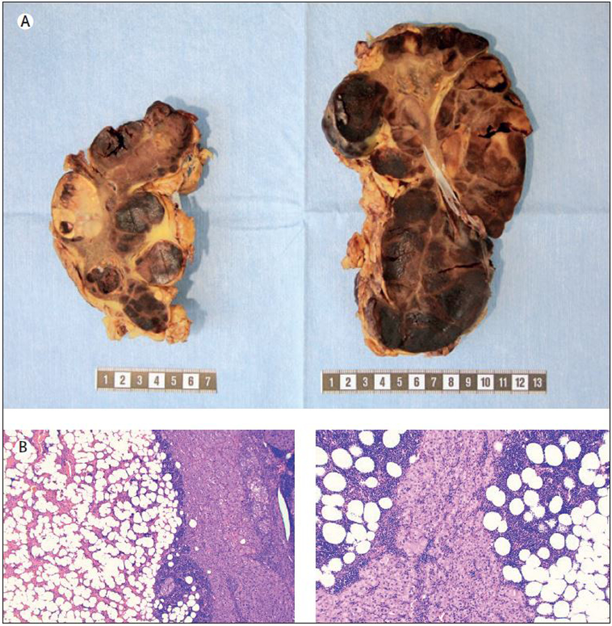
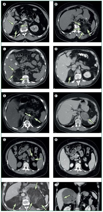
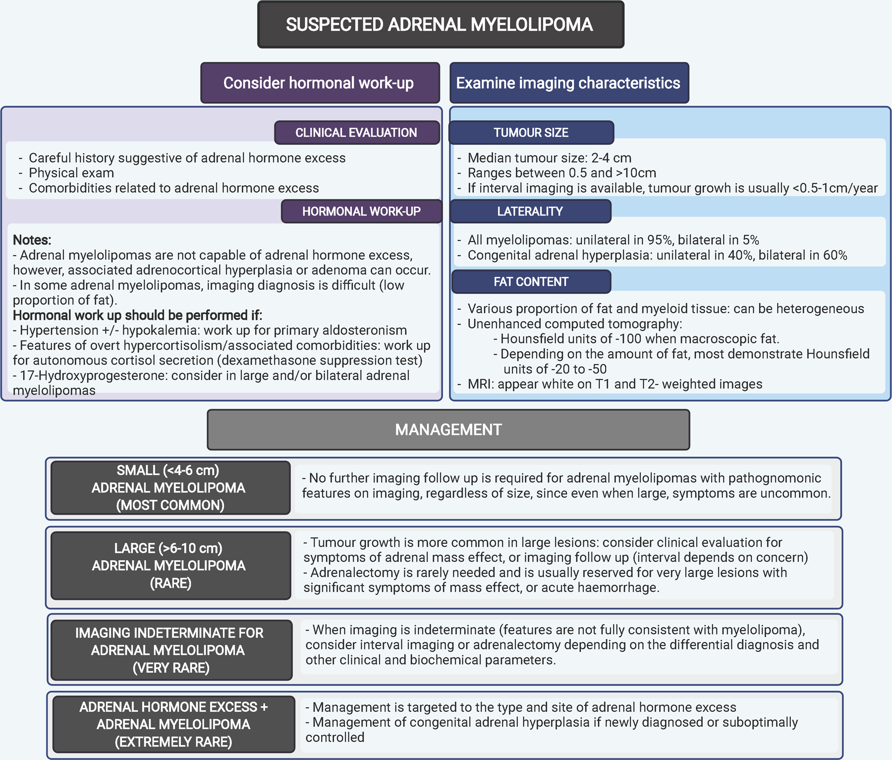
Adrenal myelolipomas are benign, lipomatous tumours with elements of myeloid cells, most of which present as adrenal incidentalomas and comprise 3·3-6·5% of all adrenal masses. Adrenal myelolipomas are usually unilateral (in 95% of cases), variable in size, most often found during midlife, and affect both sexes almost equally. On imaging, adrenal myelolipomas show pathognomonic imaging features consistent with the presence of macroscopic fat. Large adrenal myelolipomas can cause symptoms of mass effect, and can occasionally be complicated by haemorrhage. In the event of a concomitant adrenal cortical adenoma or hyperplasia, adrenal hormone excess might be detected in patients with adrenal myelolipoma. Patients with congenital adrenal hyperplasia exhibit a higher prevalence of adrenal myelolipomas than other patient groups, and are at risk of developing large and bilateral lesions. This Review discusses the pathogenesis, clinical presentation, and management of adrenal myelolipomas.


Conflict of interest statement

Declaration of interests IB is supported by the National Institute of Diabetes and Digestive and Kidney Diseases of the National Institutes of Health (NIH) USA under award K23DK121888. The views expressed are those of the authors and not necessarily those of the National Institutes of Health. IB reports consulting with Strongbridge, HRA Pharma, Corcept, CinCor, and Sparrow Pharmaceutics, and serves on the data safety board for Adrenas Therapeutics. HF has consulted for Neurocrine Biosciences, Diurnal, Roche Diagnostics International, and Adrenas Therapeutics. The other authors declare no competing interests.

Figures

Figure 1.
Figure 1.
Macroscopic and histological attributes of a bilateral adrenalectomy specimen in a patient with congenital adrenal hyperplasia. Top row: Gross image of the resected adrenals displaying multiple foci of adrenal myelolipoma (dark brownish-red areas) intermingled with hyperplastic adrenal cortical tissue (bright yellow). Metric ruler is depicted for size estimations. Bottom row: Representative photomicrographs of haematoxylin-eosin stained sections from each adrenal specimen (magnified x40 and x100 respectively) displaying myelolipoma and hyperplastic cortical tissue.
Figure 2.
Figure 2.
A-I. Transversal computed tomography (CT) images. J. Coronal CT image. A. Typical myelolipoma in the right adrenal gland (long arrow), comprising almost exclusively macroscopic fat, spreading the lateral and medial adrenal limbs apart (short arrows), extending posteriorly but demarcated from the periadrenal fat by a thin capsule. B. The myelolipoma in the left adrenal gland is dislocating the lateral adrenal limb ventrally and extends laterally to the spleen. The capsule is clearly seen (short arrows), and the myeloid component shows a cloudy appearance (long arrow). C. In the myelolipoma in the right adrenal gland, the myeloid components are seen as a nodule (long arrow) in the ventral-medial part of the tumour and as strands (small arrows) in its posterior aspects. D. In this myelolipoma in the right adrenal gland (arrow), the myeloid and fat components are mixed and are evenly spread throughout the whole tumour. E. By contrast, in this myelolipoma in the left adrenal gland, the myeloid (short arrow) and fat components (long arrow) are distinctly separated and are located in the medial and lateral part of the tumour, respectively. F. The myelolipoma in the left adrenal gland comprise almost exclusively myeloid tissue in which there is a small island of macroscopic fat in the lateral-posterior aspect of the tumour (arrow). G. CT without intravenous contrast-enhancement. In this myelolipoma in the left adrenal gland, with a similar appearance there are two small fatty islands (short arrows). H. CT with intravenous contrast-enhancement in the venous phase shows in the same tumour contrast-enhancement of the myeloid component, similar to that of the liver and spleen. I. Myelolipomas are sometimes bilateral, especially when they are large (arrows). J. Sometimes calcifications are seen in myelolipomas and they are usually small (arrow).
Figure 3.
Figure 3.
An algorithm on imaging characteristics, investigations and management of adrenal myelolipomas.

References

    1. Gierke E Über Knochenmarksgewebe in der Nebenniere. Zeiglers Beitr Pathol Anat 1905; 7: 311–24.
    1. Ebbehoj A, Li D, Kaur RJ, et al. Epidemiology of adrenal tumours in Olmsted County, Minnesota, USA: a population-based cohort study. Lancet Diabetes Endocrinol 2020; 8(11): 894–902. - PMC - PubMed
    1. Ichijo T, Ueshiba H, Nawata H, Yanase T. A nationwide survey of adrenal incidentalomas in Japan: the first report of clinical and epidemiological features. Endocr J 2020; 67(2): 141–52. - PubMed
    1. Reginelli A, Vacca G, Belfiore M, et al. Pitfalls and differential diagnosis on adrenal lesions: current concepts in CT/MR imaging: a narrative review. Gland surgery 2020; 9(6): 2331–42. - PMC - PubMed
    1. Daneshmand S, Quek ML. Adrenal myelolipoma: diagnosis and management. Urol J 2006; 3(2): 71–4. - PubMed

Publication types

Substances

Supplementary concepts