. 2021 Dec:346:113838.
doi: 10.1016/j.expneurol.2021.113838. Epub 2021 Aug 25.

Painful diabetic neuropathy leads to functional CaV3.2 expression and spontaneous activity in skin nociceptors of mice

Affiliations

Painful diabetic neuropathy leads to functional CaV3.2 expression and spontaneous activity in skin nociceptors of mice

Tal Hoffmann et al. Exp Neurol. 2021 Dec.

Abstract

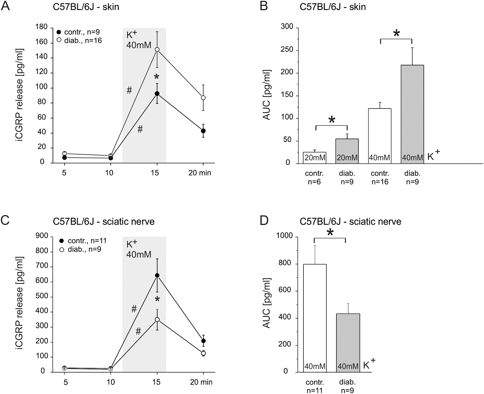
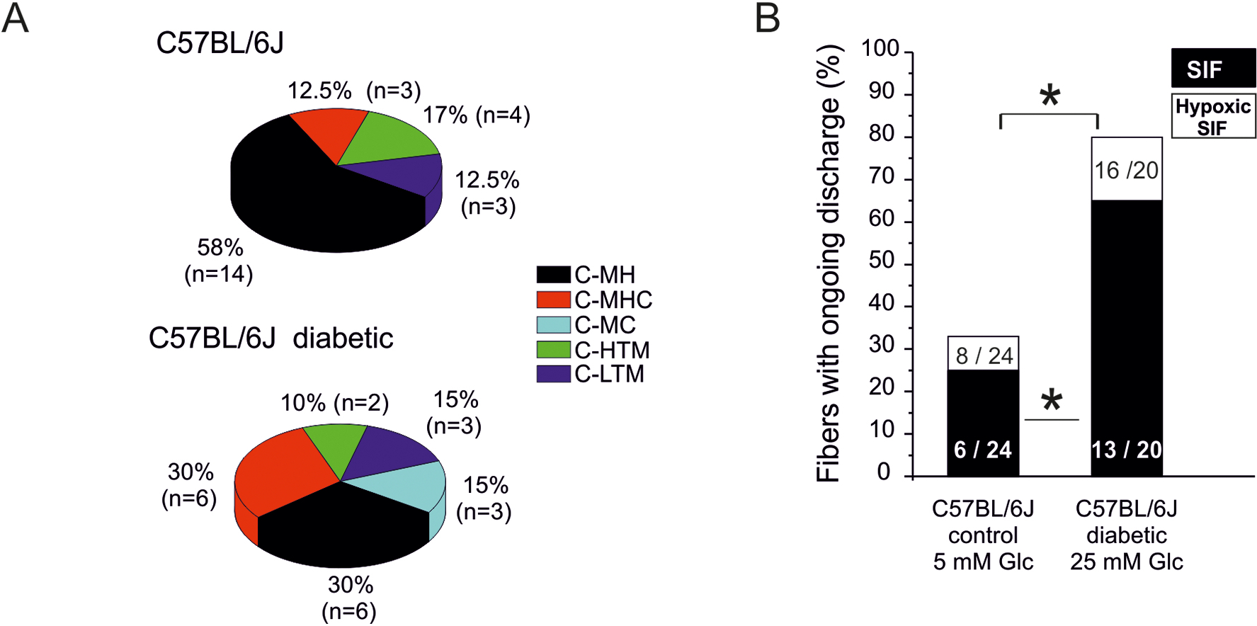
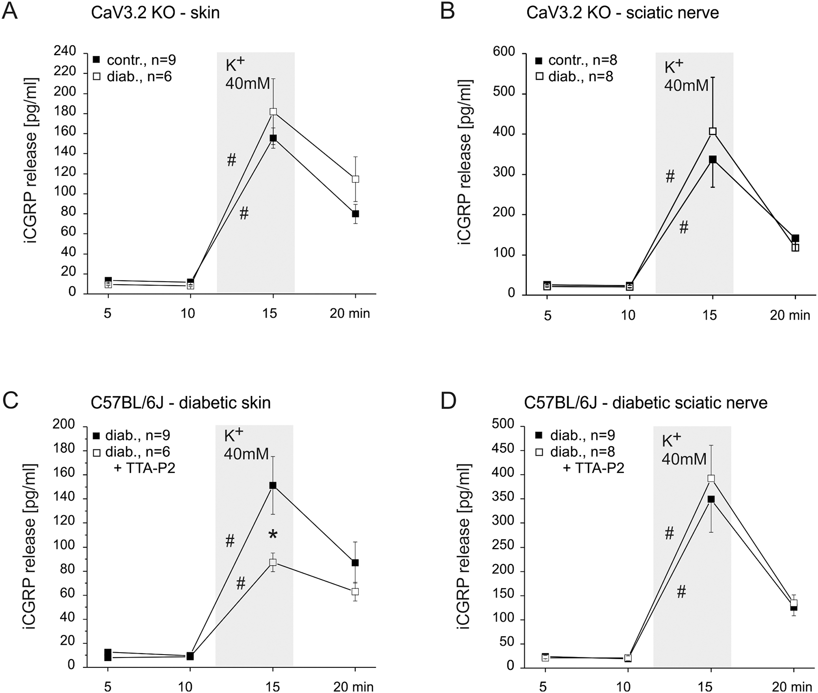
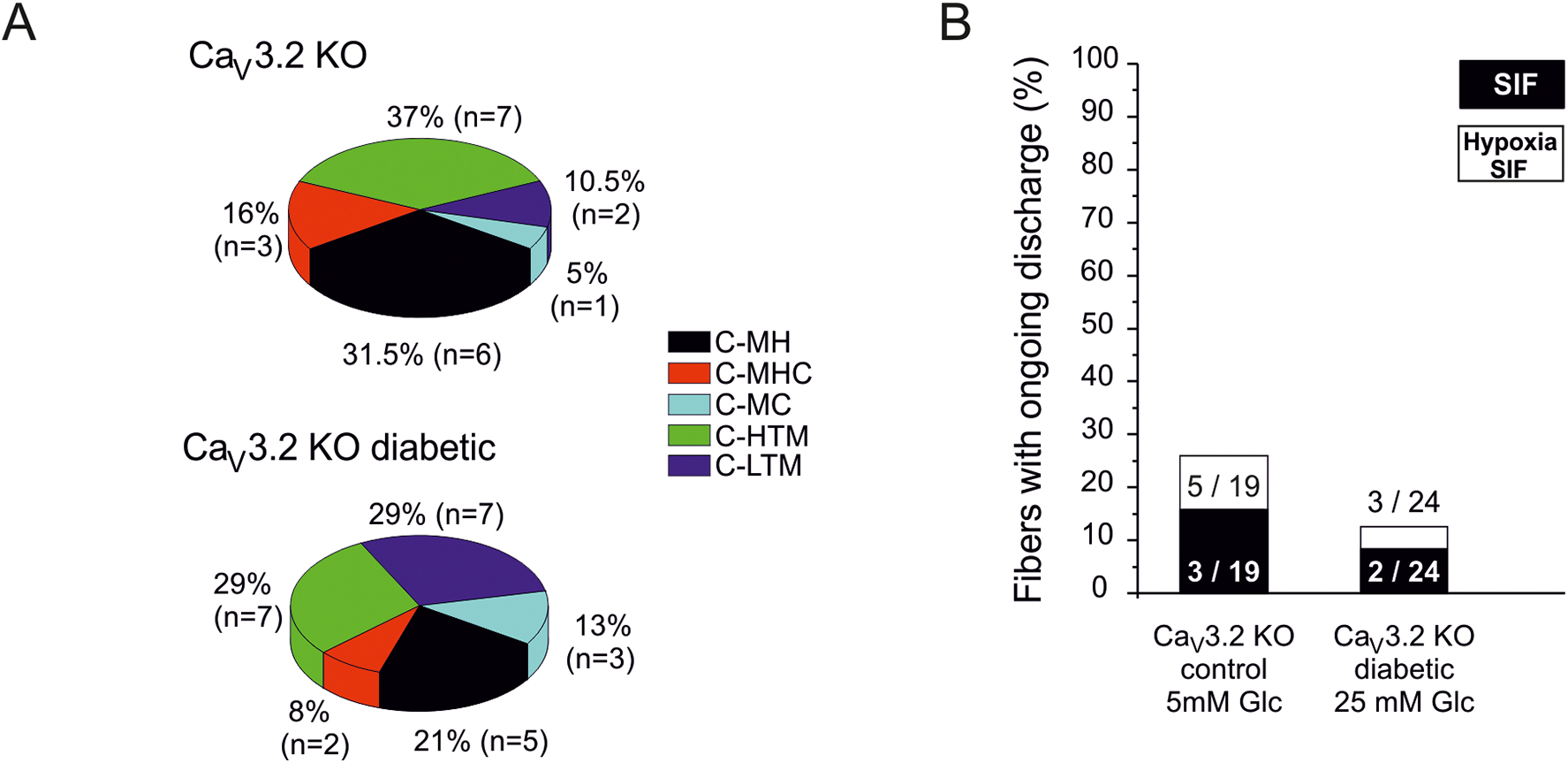
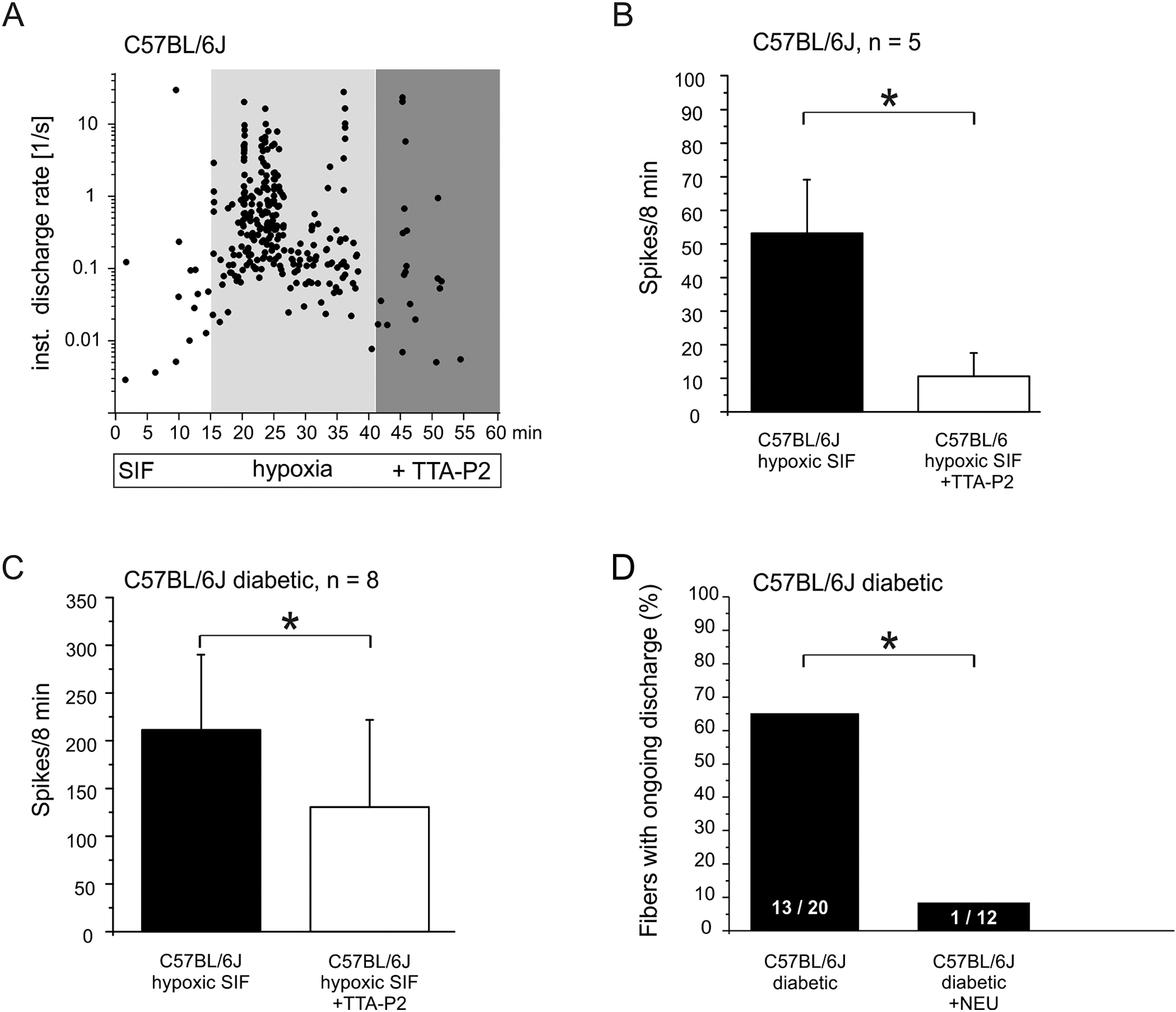
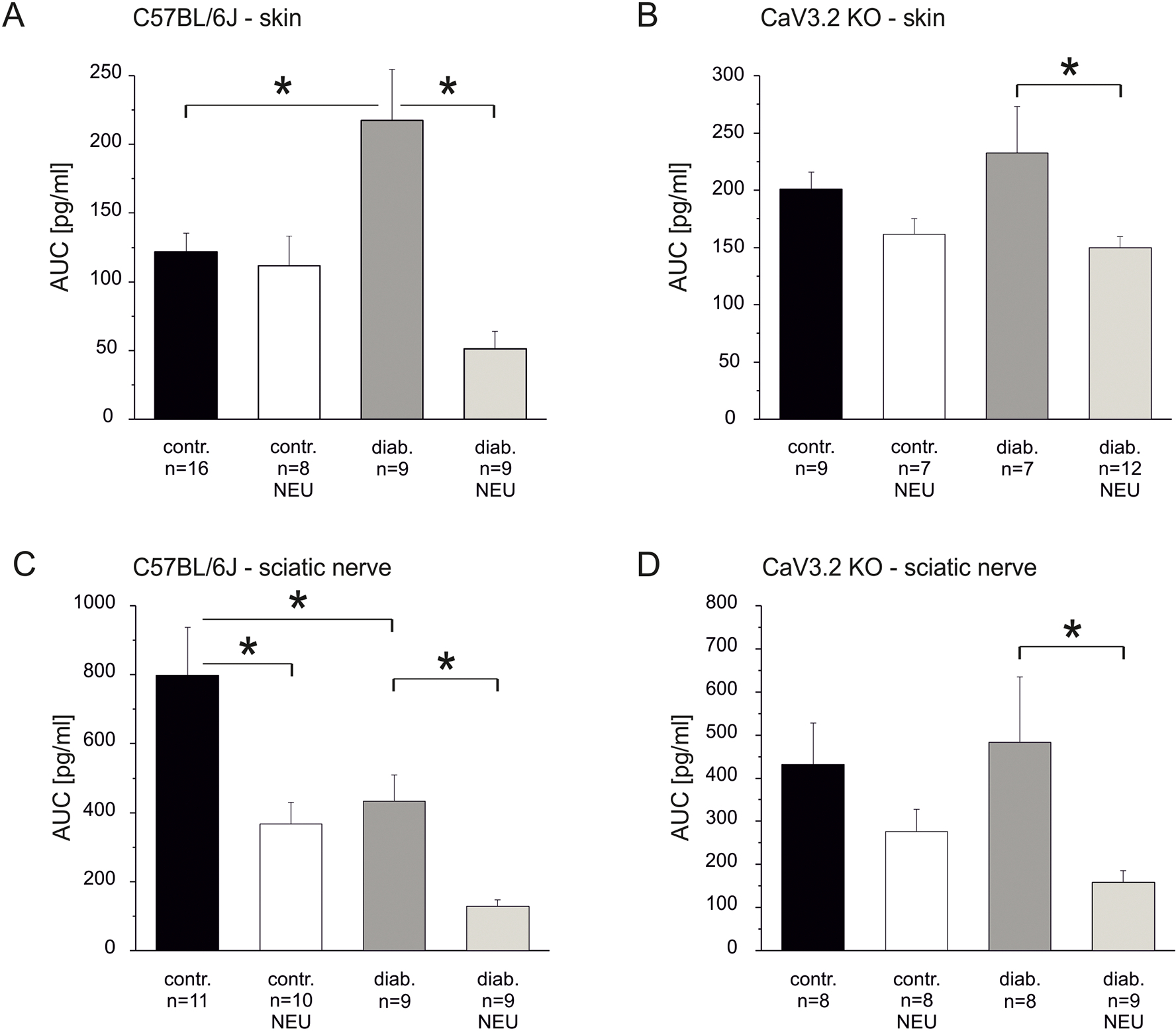
Painful diabetic neuropathy occurs in approximately 20% of diabetic patients with underlying pathomechanisms not fully understood. We evaluated the contribution of the CaV3.2 isoform of T-type calcium channel to hyperglycemia-induced changes in cutaneous sensory C-fiber functions and neuropeptide release employing the streptozotocin (STZ) diabetes model in congenic mouse strains including global knockouts (KOs). Hyperglycemia established for 3-5 weeks in male C57BL/6J mice led to major reorganizations in peripheral C-fiber functions. Unbiased electrophysiological screening of mechanosensitive single-fibers in isolated hairy hindpaw skin revealed a relative loss of (polymodal) heat sensing in favor of cold sensing. In healthy CaV3.2 KO mice both heat and cold sensitivity among the C-fibers seemed underrepresented in favor of exclusive mechanosensitivity, low-threshold in particular, which deficit became significant in the diabetic KOs. Diabetes also led to a marked increase in the incidence of spontaneous discharge activity among the C-fibers of wildtype mice, which was reduced by the specific CaV3.2 blocker TTA-P2 and largely absent in the KOs. Evaluation restricted to the peptidergic class of nerve fibers - measuring KCl-stimulated CGRP release - revealed a marked reduction in the sciatic nerve by TTA-P2 in healthy but not diabetic wildtypes, the latter showing CGRP release that was as much reduced as in healthy and, to the same extent, in diabetic CaV3.2 KOs. These data suggest that diabetes abrogates all CaV3.2 functionality in the peripheral nerve axons. In striking contrast, diabetes markedly increased the KCl-stimulated CGRP release from isolated hairy skin of wildtypes but not KO mice, and TTA-P2 reversed this increase, strongly suggesting a de novo expression of CaV3.2 in peptidergic cutaneous nerve endings which may contribute to the enhanced spontaneous activity. De-glycosylation by neuraminidase showed clear desensitizing effects, both in regard to spontaneous activity and stimulated CGRP release, but included actions independent of CaV3.2. However, as diabetes-enhanced glycosylation is decisive for intra-axonal trafficking, it may account for the substantial reorganizations of the CaV3.2 distribution. The results may strengthen the validation of CaV3.2 channel as a therapeutic target of treating painful diabetic neuropathy.

Keywords: Calcitonin gene-related peptide; Excitability; Neuropathic pain; Neuropeptide release; Sciatic nerve; T-type calcium channel; TTA-P2.


Conflict of interest statement

Conflict of interest: The authors declare that they have no conflicts of interest with the contents of this article.

Figures

Fig 1:
Fig 1:. Diabetes-induced changes in stimulated CGRP release from C57BL/6J skin and sciatic nerve.
A. KCl-evoked CGRP release from isolated hairy skin of healthy and diabetic mice. B. AUC display of the data in A plus KCl concentration dependency. C. KCl-evoked CGRP release from isolated sciatic nerve of healthy and diabetic mice. D. AUC display of the data in C. Significant increases in CGRP release over baseline and significant differences between healthy and diabetic preparations are marked with a hashtag or an asterisk based on Wilcoxon matched pairs test and ANOVA repeated measurements, respectively.
Fig. 2:
Fig. 2:. Diabetes-induced changes in C-fiber sensitivities and spontaneous activity - single-fiber recordings from hairy hindpaw skin.
A. Distribution of sensory C-fiber subclasses in healthy versus diabetic C57BL/6J mice: MH = mechano-heat, MHC = mechanoheat-cold, MC = mechano-cold, HTM = high-threshold mechano-, LTM = low-threshold mechano-sensitive. B. Incidence (% of fibers) of spontaneous discharge activity (after sensory testing) in healthy versus diabetic C57BL/6J mice and its increment during 10 min hypoxia. Significant difference between healthy and diabetic preparations is marked with an asterisk based on ANOVA repeated measurements.
Fig. 3:
Fig. 3:. Contributions of CaV3.2 to stimulated CGRP release in healthy skin and sciatic nerve.
A. KCl-evoked CGRP release from hairy skin of C57BL/6J and CaV3.2 KO mice and inhibitory effect of combined HVA calcium channel blockers nifedipine (10 μM) and ω–conotoxin (1μM). B. AUC display of data in A and no effect of the CaV3.2 blocker TTAP2 (10μM). C. KCl-evoked CGRP release from sciatic nerve of C57BL/6J and CaV3.2 KO mice. D. AUC display of the data in A and effect of the CaV3.2 blocker TTA-P2 (10μM). Significant increases in CGRP release over baseline and significant differences between experimental groups are marked with a hashtag or an asterisk based on Wilcoxon matched pairs test and ANOVA repeated measurements, respectively.
Fig. 4:
Fig. 4:. Contribution of CaV3.2 to diabetes-induced changes in stimulated CGRP release.
A. KCl-evoked CGRP release from skin of healthy and diabeticCaV3.2 KO mice. B. KCl-evoked CGRP release from sciatic nerve of healthy and diabetic CaV3.2 KO mice. C. KCl-evoked CGRP release from skin of diabetic C57BL/6J and effect of the CaV3.2 blocker TTA-P2 . D. KCl-evoked CGRP release from sciatic nerve of diabetic C57BL/6J and no effect of the CaV3.2 blocker TTA-P2. Significant increases in CGRP release over baseline and significant differences between experimental groups are marked with a hashtag an asterisk based on Wilcoxon matched pairs test and ANOVA repeated measurements, respectively.
Fig. 5:
Fig. 5:. Contribution of CaV3.2 to C-fiber sensitivities, spontaneous discharge, and diabetic changes.
A. Distribution of sensory C-fiber subclasses in in healthy versus diabetic CaV3.2 KO mice. B. Incidence (% of fibers) of spontaneous discharge activity (after sensory testing) in healthy versus diabetic CaV3.2 KO mice and its increment during 10 min hypoxia.
Fig. 6:
Fig. 6:. Pharmacological effects on spontaneous C-fiber activity of healthy versus diabetic C57BL/6J.
A. Single-fiber example of ongoing discharge intensified by hypoxia and inhibitory effect of CaV3.2 blocker TTA-P2. B. Mean ongoing discharge in final 8 min of hypoxia and subsequent inhibitory effect of TTA-P2. C. As in B but diabetic animals. D. Incidence (% of fibers) of spontaneous activity among C-fibers of diabetic skin (from Fig. 3B) versus diabetic skin after de-glycosylation pretreatment with neuraminidase. Significant differences between experimental groups are marked with an asterisk based on ANOVA repeated measurements.
Fig. 7:
Fig. 7:. Effects of de-glycosylation on stimulated CGRP release from healthy and diabetic skin and sciatic nerve.
A. KCl-evoked CGRP release from skin of healthy versus diabetic C57BL/6J mice with and without pretreatment by the de-glycosilating enzyme neuraminidase. B. KCl-evoked CGRP release from skin of healthy versus diabetic CaV3.2 KO mice with and without pretreatment by neuraminidase. C. KCl-evoked CGRP release from sciatic nerve of healthy versus diabetic C57BL/6J mice with and without pretreatment by neuraminidase. D. KCl-evoked CGRP release from sciatic nerve of healthy versus diabetic CaV3.2 KO mice with and without pretreatment by neuraminidase. Significant differences between experimental groups are marked with an asterisk based on ANOVA repeated measurements.

Similar articles

Cited by

References

    1. Abad-Rodríguez J, Díez-Revuelta N, 2015. Axon glycoprotein routing in nerve polarity, function, and repair. Trends in biochemical sciences 40, 385–396. - PubMed
    1. Amrutkar DV, Ploug KB, Olesen J, Jansen-Olesen I, 2011. Role for voltage gated calcium channels in calcitonin gene-related peptide release in the rat trigeminovascular system. Neuroscience 172, 510–517. - PubMed
    1. Andersson DA, Gentry C, Light E, Vastani N, Vallortigara J, Bierhaus A, Fleming T, Bevan S, 2013. Methylglyoxal evokes pain by stimulating TRPA1. PloS one 8, e77986. - PMC - PubMed
    1. Ayoola C, Hwang SM, Hong SJ, Rose KE, Boyd C, Bozic N, Park JY, Osuru HP, DiGruccio MR, Covey DF, Jevtovic-Todorovic V, Todorovic SM, 2014. Inhibition of CaV3.2 T-type calcium channels in peripheral sensory neurons contributes to analgesic properties of epipregnanolone. Psychopharmacology 231, 3503–3515. - PMC - PubMed
    1. Babes A, Fischer MJ, Reid G, Sauer SK, Zimmermann K, Reeh PW, 2010. Electrophysiological and neurochemical techniques to investigate sensory neurons in analgesia research. Methods Mol. Biol 617:237–59., 237–259. - PubMed

Publication types

MeSH terms

Substances