. 2021 Nov:100:108076.
doi: 10.1016/j.intimp.2021.108076. Epub 2021 Aug 17.

Matrix metalloproteinases are involved in the development of neurological complications in patients with Coronavirus disease 2019

Affiliations

Matrix metalloproteinases are involved in the development of neurological complications in patients with Coronavirus disease 2019

Mina Mohammadhosayni et al. Int Immunopharmacol. 2021 Nov.

Abstract

Background: Evidence show that Matrix metalloproteinases (MMPs) have been associated with neurological complications in the viral infections. Here in the current investigation, we intended to reveal if MMPs are potentially involved in the development of neurological symptoms in the patients with Coronavirus disease 2019 (COVID-19).

Methods: The levels of MMPs, inflammatory cytokines, chemokines, and adhesion molecules were evaluated in the serum and cerebrospinal fluid (CSF) samples from 10 COVID-19 patients with neurological syndrome (NS) and 10 COVID-19 patients lacking NS. Monocytes from the CSF samples were treated with TNF-α and the secreted levels of MMPs were determined.

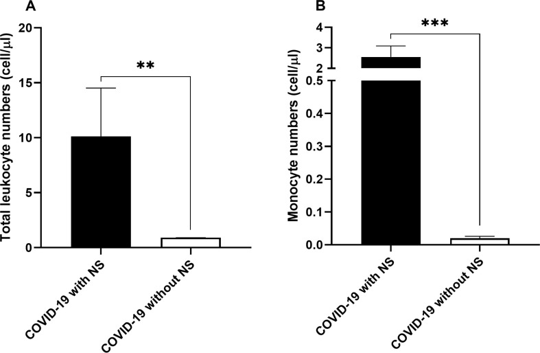
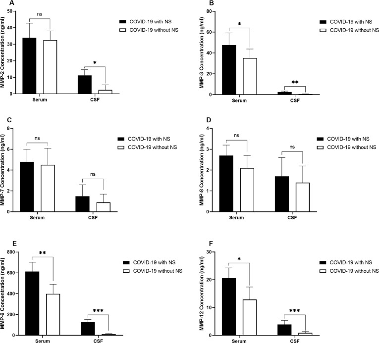
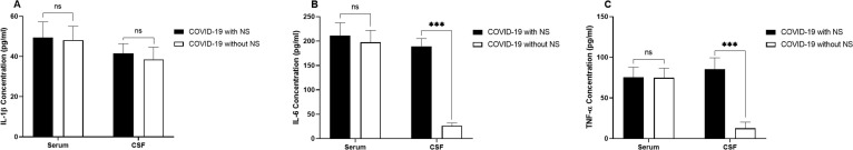
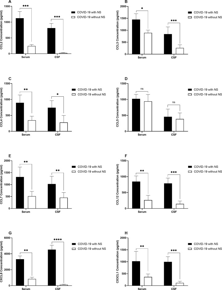
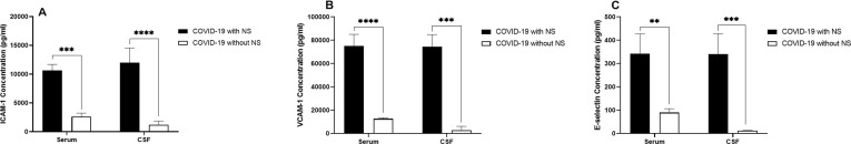
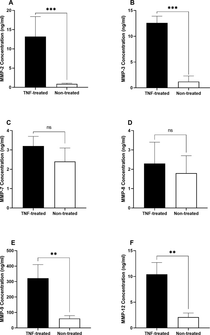
Results: The frequency of monocytes were increased in the CSF samples of COVID-19 patients with NS compared to patients without NS. Levels of inflammatory cytokines IL-1β, IL-6, and TNF-α, chemokines CCL2, CCL3, CCL4, CCL7, CCL12, CXCL8, and CX3CL1, MMPs MMP-2, MMP-3, MMP-9, and MMP-12, and adhesion molecules ICAM-1, VCAM-1, and E-selectin were significantly increased in the CSF samples of COVID-19 patients with NS compared with patients without NS. Treatment of CSF-derived monocytes obtained from COVID-19 patients with NS caused increased production of MMP-2, MMP-3, MMP-9, and MMP-12.

Conclusions: Higher levels of inflammatory cytokines might promote the expression of adhesion molecules on blood-CSF barrier (BCSFB), resulting in facilitation of monocyte recruitment. Increased levels of CSF chemokines might also help to the trafficking of monocytes to CSF. Inflammatory cytokines might enhance production of MMPs from monocytes, leading to disruption of BCSFB (and therefore further infiltration of inflammatory cells to CSF) in COVID-19 patients with NS.

Keywords: Adhesion molecule; Chemokine; Coronavirus disease 2019; Inflammatory cytokine; Matrix metalloproteinases; Neurological symptom.


Conflict of interest statement

The authors declare that they have no known competing financial interests or personal relationships that could have appeared to influence the work reported in this paper.

Figures

Fig. 1
Fig. 1
Numbers of total leukocytes (A) and monocytes (B) in the CSF samples obtained from 10 COVID-19 patients with NS and 10 COVID-19 patients without NS. The leukocyte profiling of the samples was determined by routine laboratory hematologic analyzer. The Mann-Whitney U test was used to compare the data between two groups. Data are presented by mean ± SD of the results from 10 cases in each group (NS; Neurologic syndrome, ** shows a P < 0.01 and *** shows a P < 0.001).
Fig. 2
Fig. 2
Bar graphs show the serum and CSF levels of MMP-2 (A), MMP-3 (B), MMP-7 (C), MMP-8 (D), MMP-9 (E), and MMP-12 (F) in 10 COVID-19 patients with NS and 10 COVID-19 patients without NS. The concentration of MMPs was measured using ELISA method. The Mann-Whitney U test was used to compare the data between two groups. Data are presented by mean ± SD of the results from 10 cases in each group (NS; Neurologic syndrome, ns; non-significant, * shows a P < 0.5 ** shows a P < 0.01, *** shows a P < 0.001).
Fig. 3
Fig. 3
Demonstration of the serum and CSF levels of IL-1β (A), IL-6 (B), and TNF-α (C), in 10 COVID-19 patients with NS and 10 COVID-19 cases without NS. The concentration of cytokines was measured using ELISA method. The Mann-Whitney U test was used to compare the data between two groups. Data are presented by mean ± SD of the results from 10 cases in each group (NS; Neurologic syndrome, ns; non-significant, *** shows a P < 0.001).
Fig. 4
Fig. 4
Bar graphs show the serum and CSF levels of CCL2 (A), CCL3 (B), CCL4 (C), CCL5 (D), CCL7 (E), CCL12 (F), CXCL8 (G), and CX3CL1 (H) in COVID-19 patients with NS and COVID-19 patients without NS. The concentration of chemokines was measured using ELISA method. The Mann-Whitney U test was used to compare the data between two groups. Data are presented by mean ± SD of the results from 10 cases in each group (NS; Neurologic syndrome, ns; non-significant, * shows a P < 0.5 ** shows a P < 0.01, *** shows a P < 0.001, **** shows a P < 0.0001).
Fig. 5
Fig. 5
Illustration of the serum and CSF levels of ICAM-1 (A), VCAM-1 (B), and E-selectin (C), in 1 0COVID-19 patients with NS and 10 COVID-19 cases without NS. The concentration of adhesion molecules was measured using ELISA method. The Mann-Whitney U test was used to compare the data between two groups. Data are presented by mean ± SD of the results from 10 cases in each group (NS; Neurologic syndrome, ns; non-significant, *** shows a P < 0.001).
Fig. 6
Fig. 6
Levels of MMPs in the supernatant of TNF-α treated and non-treated monocytes derived from CSF samples of COVID-19 patients with NS. Monocytes were isolated from the total leukocytes in the CSF samples by positive selection of CD14 + cells using magnetic-activated cell sorter columns. Afterwards, monocytes were incubated with 100 ng/ml Lipopolysaccharides and 5 ng/ml recombinant TNF for 72 h. The supernatant of each well was collected to measure the levels of MMPs. The concentration of MMPs was measured using ELISA method. The Mann-Whitney U test was used to compare the data between two groups. Data are presented by mean ± SD of the results from 10 cases in each group. After treating monocytes with TNF, level of MMP-2 (A), MMP-3 (B), MMP-9 (E), and MMP-12 (F) was significantly higher in comparison to untreated monocytes obtained from CSF samples of COVID-19 patients with NS. There were no significant differences in the secreted levels of MMP-7 (C) and MMP-8 (D) (TNF; Tumor necrosis factor, ns; non-significant, ** shows a P < 0.01 and *** shows a P < 0.001).

References

    1. N. Ebrahimi, S. Aslani, F. Babaie, M. Hemmatzadeh, R. Hosseinzadeh, Z. Joneidi, Z.M. Tourzani, N. Pakravan, H. Mohammadi, Recent findings on the Coronavirus disease 2019 (COVID-19); immunopathogenesis and immunotherapeutics, International immunopharmacology (2020) 107082. - PMC - PubMed
    1. Tabrizi Z.A., Khosrojerdi A., Aslani S., Hemmatzadeh M., Babaie F., Bairami A., Shomali N., Hosseinzadeh R., Safari R., Mohammadi H. Multi-facets of neutrophil extracellular trap in infectious diseases: Moving beyond immunity. Microb. Pathog. 2021;158 - PubMed
    1. Abobaker A., Raba A.A., Alzwi A. Extrapulmonary and atypical clinical presentations of COVID-19. J. Med. Virol. 2020;92(11):2458–2464. - PMC - PubMed
    1. Helms J., Kremer S., Merdji H., Clere-Jehl R., Schenck M., Kummerlen C., Collange O., Boulay C., Fafi-Kremer S., Ohana M. Neurologic features in severe SARS-CoV-2 infection. N. Engl. J. Med. 2020;382(23):2268–2270. - PMC - PubMed
    1. Moriguchi T., Harii N., Goto J., Harada D., Sugawara H., Takamino J., Ueno M., Sakata H., Kondo K., Myose N. A first case of meningitis/encephalitis associated with SARS-Coronavirus-2. International Journal of Infectious Diseases. 2020;94:55–58. - PMC - PubMed