

Critical Review of Biodegradable and Bioactive Polymer Composites for Bone Tissue Engineering and Drug Delivery Applications
- PMID: 34451161
- PMCID: PMC8399915
- DOI: 10.3390/polym13162623
Critical Review of Biodegradable and Bioactive Polymer Composites for Bone Tissue Engineering and Drug Delivery Applications
Abstract
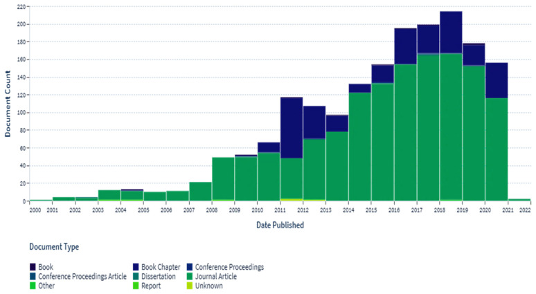
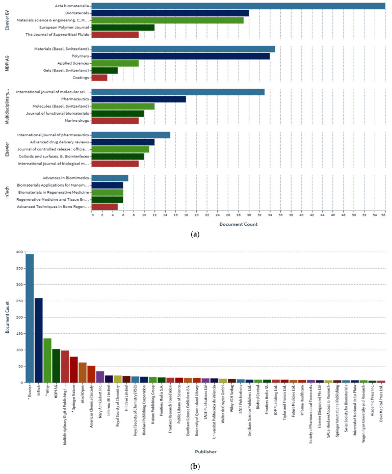
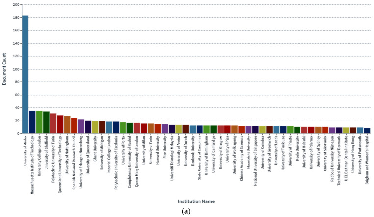
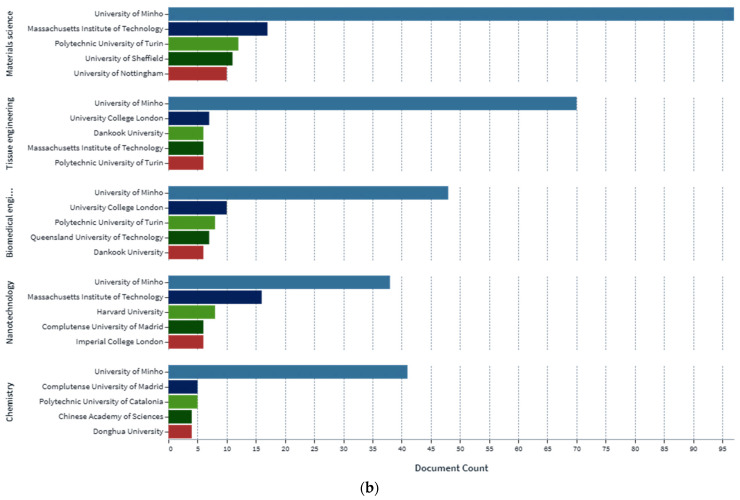
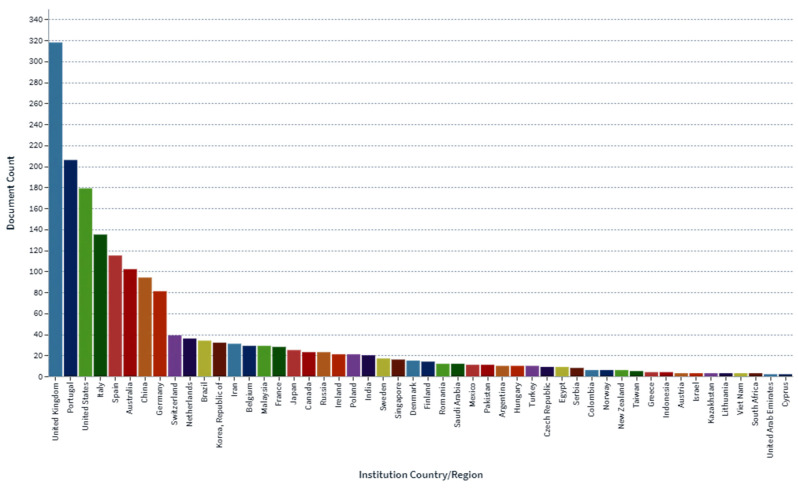
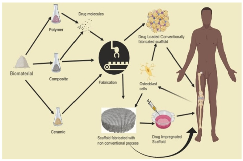
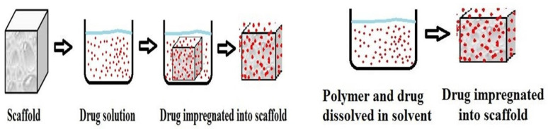
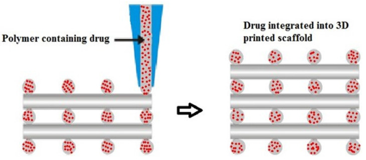
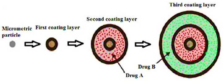
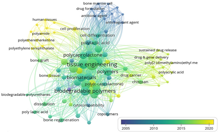
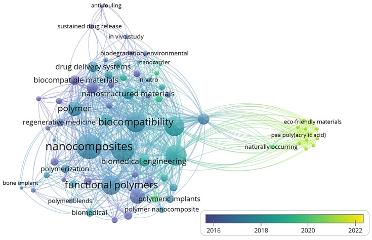
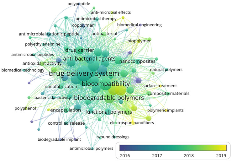
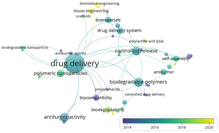
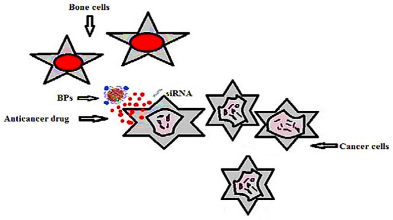
In the determination of the bioavailability of drugs administered orally, the drugs' solubility and permeability play a crucial role. For absorption of drug molecules and production of a pharmacological response, solubility is an important parameter that defines the concentration of the drug in systemic circulation. It is a challenging task to improve the oral bioavailability of drugs that have poor water solubility. Most drug molecules are either poorly soluble or insoluble in aqueous environments. Polymer nanocomposites are combinations of two or more different materials that possess unique characteristics and are fused together with sufficient energy in such a manner that the resultant material will have the best properties of both materials. These polymeric materials (biodegradable and other naturally bioactive polymers) are comprised of nanosized particles in a composition of other materials. A systematic search was carried out on Web of Science and SCOPUS using different keywords, and 485 records were found. After the screening and eligibility process, 88 journal articles were found to be eligible, and hence selected to be reviewed and analyzed. Biocompatible and biodegradable materials have emerged in the manufacture of therapeutic and pharmacologic devices, such as impermanent implantation and 3D scaffolds for tissue regeneration and biomedical applications. Substantial effort has been made in the usage of bio-based polymers for potential pharmacologic and biomedical purposes, including targeted deliveries and drug carriers for regulated drug release. These implementations necessitate unique physicochemical and pharmacokinetic, microbiological, metabolic, and degradation characteristics of the materials in order to provide prolific therapeutic treatments. As a result, a broadly diverse spectrum of natural or artificially synthesized polymers capable of enzymatic hydrolysis, hydrolyzing, or enzyme decomposition are being explored for biomedical purposes. This summary examines the contemporary status of biodegradable naturally and synthetically derived polymers for biomedical fields, such as tissue engineering, regenerative medicine, bioengineering, targeted drug discovery and delivery, implantation, and wound repair and healing. This review presents an insight into a number of the commonly used tissue engineering applications, including drug delivery carrier systems, demonstrated in the recent findings. Due to the inherent remarkable properties of biodegradable and bioactive polymers, such as their antimicrobial, antitumor, anti-inflammatory, and anticancer activities, certain materials have gained significant interest in recent years. These systems are also actively being researched to improve therapeutic activity and mitigate adverse consequences. In this article, we also present the main drug delivery systems reported in the literature and the main methods available to impregnate the polymeric scaffolds with drugs, their properties, and their respective benefits for tissue engineering.
Keywords: anticancer activity; antimicrobial properties; biodegradable polymers; drug delivery; natural bioactive polymers; polymeric scaffolds; tissue engineering.
Conflict of interest statement
The authors declare no conflict of interest.
Figures

Similar articles
-
Current development of biodegradable polymeric materials for biomedical applications.Drug Des Devel Ther. 2018 Sep 24;12:3117-3145. doi: 10.2147/DDDT.S165440. eCollection 2018. Drug Des Devel Ther. 2018. PMID: 30288019 Free PMC article. Review.
-
Biological evaluation of preceramic organosilicon polymers for various healthcare and biomedical engineering applications: A review.J Biomed Mater Res B Appl Biomater. 2021 May;109(5):744-764. doi: 10.1002/jbm.b.34740. Epub 2020 Oct 19. J Biomed Mater Res B Appl Biomater. 2021. PMID: 33075186 Review.
-
Incorporation of essential oils in polymeric films for biomedical applications.Int J Biol Macromol. 2024 Jun;269(Pt 1):132108. doi: 10.1016/j.ijbiomac.2024.132108. Epub 2024 May 6. Int J Biol Macromol. 2024. PMID: 38710258 Review.
-
Pharmaceutical, biomedical and ophthalmic applications of biodegradable polymers (BDPs): literature and patent review.Pharm Dev Technol. 2022 Mar;27(3):341-356. doi: 10.1080/10837450.2022.2055063. Epub 2022 Apr 3. Pharm Dev Technol. 2022. PMID: 35297285 Review.
-
Biodegradable Polymers as the Pivotal Player in the Design of Tissue Engineering Scaffolds.Adv Healthc Mater. 2020 Jul;9(13):e1901358. doi: 10.1002/adhm.201901358. Epub 2020 May 19. Adv Healthc Mater. 2020. PMID: 32424996 Review.
Cited by
-
Impact of Process Variables of Acetone Vapor Jet Drilling on Surface Roughness and Circularity of 3D-Printed ABS Parts: Fabrication and Studies on Thermal, Morphological, and Chemical Characterizations.Polymers (Basel). 2022 Mar 28;14(7):1367. doi: 10.3390/polym14071367. Polymers (Basel). 2022. PMID: 35406241 Free PMC article.
-
Effects of Elevated Temperature on the Residual Behavior of Concrete Containing Marble Dust and Foundry Sand.Materials (Basel). 2022 May 19;15(10):3632. doi: 10.3390/ma15103632. Materials (Basel). 2022. PMID: 35629658 Free PMC article.
-
Recent Trends and Developments in Conducting Polymer Nanocomposites for Multifunctional Applications.Polymers (Basel). 2021 Aug 28;13(17):2898. doi: 10.3390/polym13172898. Polymers (Basel). 2021. PMID: 34502938 Free PMC article. Review.
-
Multi-omics insights into bone tissue injury and healing: bridging orthopedic trauma and regenerative medicine.Burns Trauma. 2025 May 28;13:tkaf019. doi: 10.1093/burnst/tkaf019. eCollection 2025. Burns Trauma. 2025. PMID: 40438296 Free PMC article. Review.
-
Poly(propylene fumarate)/hydroxyapatite nanocomposite/black phosphorus nanosheet phosphate composites for enhanced bone repair.J Orthop Surg Res. 2025 Jul 30;20(1):721. doi: 10.1186/s13018-025-06028-z. J Orthop Surg Res. 2025. PMID: 40739523 Free PMC article.
References
-
- Bagde A., Kuthe A.M., Quazi S., Gupta V., Jaiswal S., Jyothilal S., Lande N., Nagdeve S. State of the Art Technology for Bone Tissue Engineering and Drug Delivery. IRBM. 2019;40:133–144. doi: 10.1016/j.irbm.2019.03.001. - DOI
-
- Th S., Reis R. Functional Marine Biomaterials: Properties and Applications. Woodhead Publishing; Sawston, Cambridge, UK: 2015. Drug delivery systems and cartilage tissue engineering scaffolding using marine-derived products; pp. 123–136.
Publication types
LinkOut - more resources
Full Text Sources
Research Materials
