Review
. 2021 Aug 23;13(16):2833.
doi: 10.3390/polym13162833.

Biomedical Applications of Antiviral Nanohybrid Materials Relating to the COVID-19 Pandemic and Other Viral Crises

Affiliations
Review

Biomedical Applications of Antiviral Nanohybrid Materials Relating to the COVID-19 Pandemic and Other Viral Crises

Shahin Homaeigohar et al. Polymers (Basel). .

Abstract

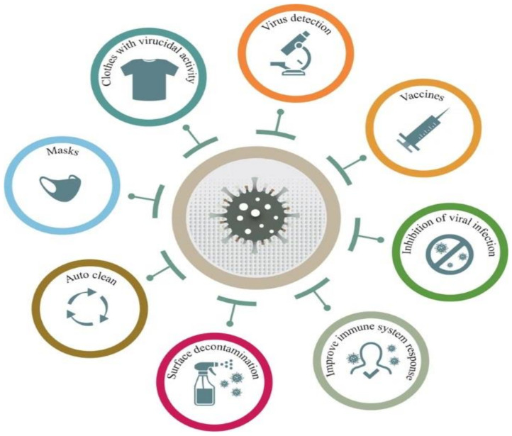
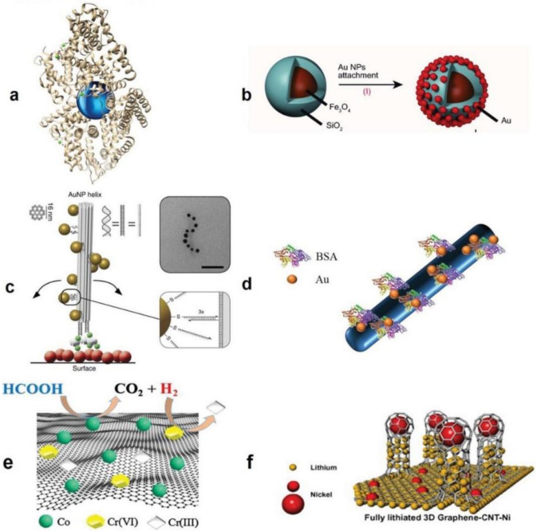
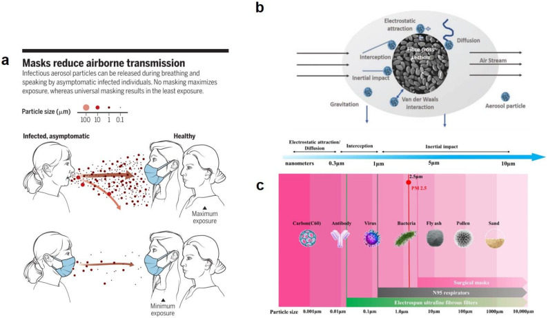
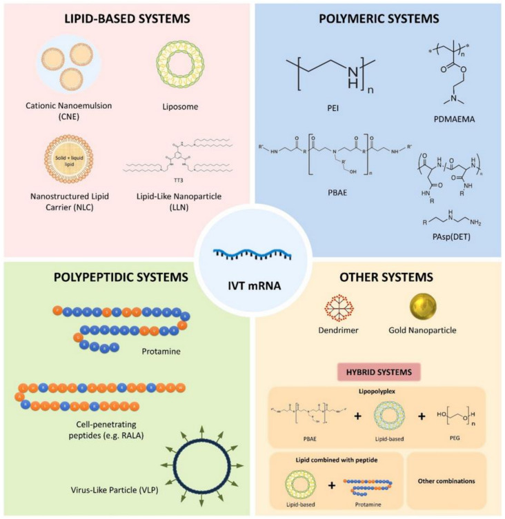
The COVID-19 pandemic has driven a global research to uncover novel, effective therapeutical and diagnosis approaches. In addition, control of spread of infection has been targeted through development of preventive tools and measures. In this regard, nanomaterials, particularly, those combining two or even several constituting materials possessing dissimilar physicochemical (or even biological) properties, i.e., nanohybrid materials play a significant role. Nanoparticulate nanohybrids have gained a widespread reputation for prevention of viral crises, thanks to their promising antimicrobial properties as well as their potential to act as a carrier for vaccines. On the other hand, they can perform well as a photo-driven killer for viruses when they release reactive oxygen species (ROS) or photothermally damage the virus membrane. The nanofibers can also play a crucial protective role when integrated into face masks and personal protective equipment, particularly as hybridized with antiviral nanoparticles. In this draft, we review the antiviral nanohybrids that could potentially be applied to control, diagnose, and treat the consequences of COVID-19 pandemic. Considering the short age of this health problem, trivially the relevant technologies are not that many and are handful. Therefore, still progressing, older technologies with antiviral potential are also included and discussed. To conclude, nanohybrid nanomaterials with their high engineering potential and ability to inactivate pathogens including viruses will contribute decisively to the future of nanomedicine tackling the current and future pandemics.

Keywords: COVID-19; biomedical application; nanocomposite; nanohybrid.


Conflict of interest statement

The authors declare no conflict of interest.

Figures

Figure 1
Figure 1
Biomedical applications of nanomaterials against the COVID-19 pandemic. Reproduced with permission [7] under a Creative Commons Attribution 4.0 International License.
Figure 2
Figure 2
Nanohybrid materials in different dimensionalities, forms, and configurations. (a) A 0D bionanohybrid comprised of a plasmonic nanoparticle coupled with a protein, i.e., bovine serum albumin (BSA). (b) A 0D superparamagnetic core-shell nanohybrid composed of Au nanoparticles/Fe3O4@SiO2 developed via chemical immobilization of Au nanoparticles on the amino group functionalized SiO2 surface. (c) A 1D nanohybrid made of Au nanoparticle/DNA; in this system the Au nanoparticles (10 nm) are organized as a secondary left-handed helix located on the DNA origami bundle. The bundle per se consists of 24 parallel double helices. The magnified zone indicates that the thiolated ssDNA functionalized Au nanoparticles are coupled to the DNA origami. Next, functionalization of the DNA origami by the biotin groups (green) enables its attachment to a surface coated with BSA–biotin–neutravidin (red, green, and grey, respectively). Transmission electron microscopy (TEM) image exhibits a nanohelix mounted on a carbon-deposited grid (the scale bar represents 50 nm). (d) A 1D nanohybrid based on protein functionalized polymer nanofibers whereon Au nanoparticles are coupled by the protein ligands. (e) A 2D nanohybrid composed of cobalt-reduced graphene oxide (Co-rGO) (as shown in the figure, hexavalent chromium (Cr(VI)) is reduced by the nanohybrid). (f) A 3D hierarchical nanohybrid made of graphene–carbon nanotube–nickel (G-CNT-Ni), meant to operate as an anode material in the fully lithiated state. Reproduced with permission [8] Copyright 2018, Wiley-VCH.
Figure 3
Figure 3
(a) airborne transmission of infectious aerosol particles is notably reduced by wearing masks. Reproduced with permission [17], Copyright 2020, Science. (b) Operation mechanisms of fibrous face masks depending on the size of the filtrate. (c) size (length) scale of different particles available in nature and the respective filtration means including surgical masks, N95 respirators, and electrospun fibrous filters. Reproduced with permission [18], Copyright 2021, Elsevier.
Figure 4
Figure 4
The as-yet studied mRNA vaccine nanocarriers. Reproduced with permission [38].
Figure 5
Figure 5
(a) Schematic illustrates the solar light driven photocatalysis (Ehv and Eg are the solar light photon energy and band gap energy, respectively, and, CB and VB are the conduction and valence band, respectively. R and D are the electron acceptor and electron donor, respectively. Reproduced with permission [68], Copyright 2017, Elsevier. (b) The Ag-CuFe2O4@WO3 nanohybrid nanoparticles kill E. coli within the course of a 60 h incubation period upon exposure to UV light. The density of the bacteria colonies imaged in the absence of the nanohybrid nanoparticles (c), and in the presence of CuFe2O4@WO3 (d) and Ag-CuFe2O4@WO3 nanoparticles (e). Reproduced with permission [67].
Figure 6
Figure 6
(a) Schematic illustration of the optical/electrochemical detection process of the Influenza virus based on implementation of plasmonic nanocomposites and quantum dots. Reproduced with permission [94], Copyright 2021, American Chemical Society. The improvement of the PCR efficiency (nano-PCR based on pan-type primers) through inclusion of GO-AuNPs at different concentrations for the purpose of quantification of FMDV O-type (b) and FMDV A-type (c). Reproduced with permission [95].
Figure 7
Figure 7
(a) The mechanism of photosensitization as described through a Modified Jablonski Diagram. Upon light irradiation, a photosensitizer (PS) molecule undergoes a transition from the ground singlet state (S0) to a provoked singlet state (S1). At S1 the molecule might experience intersystem crossing towards an excited triplet state (T1). Subsequently, it either creates radicals (Type I process) or pass its energy on a molecular oxygen (3O2), forming singlet oxygen (1O2), i.e., the main cytotoxic substance taking part in PDT. In the diagram, ns, μs, nm, and eV represent nanoseconds, microseconds, nanometers, and electron volts, respectively. (b) The schematic illustration of PDT for cancer therapy, where a PS is distributed topically or systemically within body. Over time, PS is selectively accumulated in the tumor and then by irradiation is excited. Subjected to molecular oxygen, a photochemical process is carried out that leads to formation of singlet oxygen (1O2). Severe destruction of cellular macromolecules causes the cell death of tumor through a necrotic, autophagic, or apoptotic process, that is complemented with inflammation. An acute local inflammatory process eliminates the dead cells and restores normal tissue homeostasis. Reproduced with permission [130], Copyright 2011, American Cancer Society.
Figure 8
Figure 8
Schematic demonstration of the fabrication process of ZnO–Ag/PMMA nanofiber: (a) hydrothermal synthesis of ZnO nanorods. The inset image represents their morphology, (b) production of the electrospinning solution through addition of ZnO nanorods and PMMA to the AgNO3/DMF solution wherein Ag nanoparticles form via in situ reduction of the Ag salt, (c) creation of the ZnO–Ag/PMMA nanofibers through electrospinning, (d) the mats comprising the ZnO-Ag/PMMA nanofibers are employed as a protective clothing, and offer antimicrobial (e), photocatalytic (f), and sensing (g) properties. Reproduced with permission [144], Copyright 2021, American Chemical Society. TEM images show how the Ag nanoparticles adversely affect the Ad3 particles at different intervals. (h) Control sample in the absence of Ag nanoparticles; the samples treated with Ag nanoparticles (50 μg/mL) for 30 min (i), 90 min (j) and 120 min (k). As clearly observed, the capsid and the whole virus particle have been notably destructed in the presence of the nanoparticles. Reproduced with permission [158] Copyright 2013, Elsevier.
Figure 9
Figure 9
Schematic illustration of: (a) the antiviral nanohybrid coated surgical masks, (b) the dual-channel spray coating process on the nonwoven fibers of the surgical mask, whereby the Cu nanoparticle suspension and shellac are mixed and sprayed via the compressed N2 air channel, and (c) the solar light driven inactivation of the viruses available in the respiratory droplets. (d) Camera image of the photoactive antiviral mask (PAM). (e) SEM images show the morphology of propylene nonwoven fibers (left) in the commercial surgical mask and the nanohybrid-coated nonwoven fibers in the same mask (right). (Scale bar represents 10 μm). (f) Camera images of a dyed water droplet deposited on the uncoated mask and PAM after one hour. Reproduced with permission [154], Copyright 2021, American Chemical Society. (g) Schematic illustration of likely interactions between SARS-CoV-2 and graphene (GR) and GR nanohybrids comprising GR-metal ion, leading to virus inactivation on the coated surfaces. Reproduced with permission [167], Copyright 2020, Elsevier.

Similar articles

Cited by

References

    1. Weiss C., Carriere M., Fusco L., Capua I., Regla-Nava J.A., Pasquali M., Scott J.A., Vitale F., Unal M.A., Mattevi C. Toward nanotechnology-enabled approaches against the COVID-19 pandemic. ACS Nano. 2020;14:6383–6406. doi: 10.1021/acsnano.0c03697. - DOI - PubMed
    1. Lovato A., De Filippis C. Clinical presentation of COVID-19: A systematic review focusing on upper airway symptoms. Ear Nose Throat J. 2020;99:569–576. doi: 10.1177/0145561320920762. - DOI - PubMed
    1. Bansal M. Cardiovascular disease and COVID-19. Diabetes Metab. Syndr. Clin. Res. Rev. 2020;14:247–250. doi: 10.1016/j.dsx.2020.03.013. - DOI - PMC - PubMed
    1. Rabb H. Kidney diseases in the time of COVID-19: Major challenges to patient care. J. Clin. Investig. 2020;130:2749–2751. doi: 10.1172/JCI138871. - DOI - PMC - PubMed
    1. Seah I., Agrawal R. Can the coronavirus disease 2019 (COVID-19) affect the eyes? A review of coronaviruses and ocular implications in humans and animals. Ocul. Immunol. Inflamm. 2020;28:391–395. doi: 10.1080/09273948.2020.1738501. - DOI - PMC - PubMed

LinkOut - more resources