Review
. 2021 Jul 31;10(8):973.
doi: 10.3390/pathogens10080973.

The Structural Biology of Eastern Equine Encephalitis Virus, an Emerging Viral Threat

Affiliations
Review

The Structural Biology of Eastern Equine Encephalitis Virus, an Emerging Viral Threat

S Saif Hasan et al. Pathogens. .

Abstract

Alphaviruses are arboviruses that cause arthritis and encephalitis in humans. Eastern Equine Encephalitis Virus (EEEV) is a mosquito-transmitted alphavirus that is implicated in severe encephalitis in humans with high mortality. However, limited insights are available into the fundamental biology of EEEV and residue-level details of its interactions with host proteins. In recent years, outbreaks of EEEV have been reported mainly in the United States, raising concerns about public safety. This review article summarizes recent advances in the structural biology of EEEV based mainly on single-particle cryogenic electron microscopy (cryoEM) structures. Together with functional analyses of EEEV and related alphaviruses, these structural investigations provide clues to how EEEV interacts with host proteins, which may open avenues for the development of therapeutics.

Keywords: alphavirus; antibody; assembly; eastern equine encephalitis virus; structure.


Conflict of interest statement

The authors declare no competing financial interest.

Figures

Figure 1
Figure 1
Entry and replication cycle of alphaviruses. Entry and disassembly steps are highlighted in red ((1) to (4)), whereas progeny virus assembly and exit are from step (5) to (7). (Step 1) Receptor-mediated endocytosis allows alphavirus particles to enter cells (particles shown in blue, cyan). (Step 2) Viral and endosomal membranes undergo fusion upon acidification of the endosome lumen (pink). (Step 3) Cytosolic release of the nucleocapsid core (yellow). (Step 4) Disintegration of the core and release of the viral RNA genome (vRNA, red line). (Step 5) Cytosolic progeny RNA genome synthesis and nucleocapsid core assembly occur in the cytosol, whereas envelope proteins are synthesized in secretory ER and Golgi network (Step 6). (Step 7) Final assembly of alphavirus progeny particles involves plasma membrane. Figure taken from [33].
Figure 2
Figure 2
Structure of EEEV. (a) Surface representation of EEEV using radial coloring. An internal section of the cryoEM map is shown on the right (black dotted lines highlight lipid bilayer). (b) Resolution range and distribution in the cryoEM map highlighting prominent features such as envelope proteins and capsid protein (scale at bottom). The RNA genome (gray) was not included in this analysis. Gray arrows show directions of icosahedral symmetry axes for reference. Color code for boxes: red, E1 ecto-domain; black, capsid proteins in vicinity of icosahedral 5-fold axis; “*”, E2 ecto-domain. (ce) Organization of E1–E2 trimeric spike ecto-domain shown in (c) in a radial orientation and (d) side-view rotated by 90° from (c). For clarity, E1 and E2 ecto-domains are shown in different representations as ribbon and surface, respectively. (e) Domain organization of E1 and E2 ecto-domains. The color codes as in (c,d). “F” represents the fusion loop. Figure taken from [33].
Figure 3
Figure 3
Electrostatic interactions in SINV-EEEV E1–E2. (a) Complementary charged surfaces in E1 (cyan) and E2 (pink) ecto-domain interface. Color code, acidic in red and basic in blue. (bd) Basic residues shown as sticks in the trimeric E2–E2 interface (pink). (b) Trimeric spike along 3-fold axis. Black box highlights the basic interfacial residues, which are magnified in (c). (d) Four basic residues from one E2 ecto-domain are labelled. Figure taken from [33].
Figure 4
Figure 4
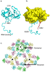
Structure of alphavirus capsid protein. (a) Capsid protein from SINV-EEEV. The RBS and NTD form an extended structure that is associated with the compact chymotrypsin-like CTD. (b) Structural superposition of SINV-EEEV and SINV capsid CTD (yellow surface) demonstrates difference in location of NTD residues between capsid of EEEV (cyan) and SINV (red). (c) In the internal capsid layer, pentamers and hexamers are arranged at the icosahedral 5- and 2-fold axes, respectively. These provide major stabilizing capsid–capsid contacts, whereas no interactions are observed near the 3-fold. In (a,b), coordinates for capsid protein (PDB ID 6MX7 for SINV-EEEV, 1SVP for SINV) were downloaded from PDB and figures were generated in PyMol (www.pymol.org, (accessed on 1 June 2021), version 1.8.6.0). Panel (c) taken from [33].
Figure 5
Figure 5
HS binding residues in E2 of SINV-EEEV. (a) The Cα-atoms of HS binding residues (Lys71, red; Lys74, yellow; Lys77, blue) shown as spheres, whereas the E2 ectodomain is shown in a surface representation (pink). (b) A rotated side-view shows one linear triad highlighted in a black oval. Figure taken from [33].
Figure 6
Figure 6
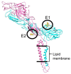
Glycosylation in EEEV E1-E2 proteins. The glycan groups are highlighted as sticks in yellow-red and are encircled for clarity. Color code, E1, cyan, E2, pink. Figure taken from [33].
Figure 7
Figure 7
CryoEM structures of Fabs in complex with SINV-EEEV chimeric particles. The footprints of the Fabs are highlighted in yellow on the trimeric spike. Figure taken from [33].
Figure 8
Figure 8
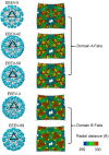
The role of steric limitations on Fab occupancy in alphaviruses. Color code: E1-E2, gray-yellow; Fab, blue; Fab quasi-2-fold axis, black dotted line. Tangential binding allows higher occupancy of domain A Fabs (upper left) unlike radial binding that is limited by clashes (upper right). In domain B Fabs, tangential binding is limited by clashes between Fabs bound to neighboring spikes and potentially neighboring E1–E2 proteins (lower left). High occupancy in domain B Fabs is favored in radial binding (lower right). Figure taken from [33].

References

    1. Strauss J.H., Strauss E.G. The alphaviruses: Gene expression, replication, and evolution. Microbiol. Rev. 1994;58:491–562. doi: 10.1128/mr.58.3.491-562.1994. - DOI - PMC - PubMed
    1. Suhrbier A., Jaffar-Bandjee M.C., Gasque P. Arthritogenic alphaviruses—An overview. Nat. Rev. Rheumatol. 2012;8:420–429. doi: 10.1038/nrrheum.2012.64. - DOI - PubMed
    1. Levi L.I., Vignuzzi M. Arthritogenic alphaviruses: A worldwide emerging threat? Microorganisms. 2019;7:133. doi: 10.3390/microorganisms7050133. - DOI - PMC - PubMed
    1. Lwande O.W., Obanda V., Bucht G., Mosomtai G., Otieno V., Ahlm C., Evander M. Global emergence of alphaviruses that cause arthritis in humans. Infect. Ecol. Epidemiol. 2015;5:29853. doi: 10.3402/iee.v5.29853. - DOI - PMC - PubMed
    1. Rulli N.E., Melton J., Wilmes A., Ewart G., Mahalingam S. The molecular and cellular aspects of arthritis due to alphavirus infections: Lesson learned from Ross River virus. Ann. N. Y. Acad. Sci. 2007;1102:96–108. doi: 10.1196/annals.1408.007. - DOI - PubMed

LinkOut - more resources