. 2021 Jul 23;10(8):1515.
doi: 10.3390/plants10081515.

Ultrastructural Alterations in Cells of Sunflower Linear Glandular Trichomes during Maturation

Affiliations

Ultrastructural Alterations in Cells of Sunflower Linear Glandular Trichomes during Maturation

Evelyn Amrehn et al. Plants (Basel). .

Abstract

Sunflower and related taxa are known to possess a characteristic type of multicellular uniseriate trichome which produces sesquiterpenes and flavonoids of yet unknown function for this plant. Contrary to the metabolic profile, the cytological development and ultrastructural rearrangements during the biosynthetic activity of the trichome have not been studied in detail so far. Light, fluorescence and transmission electron microscopy were employed to investigate the functional structure of different trichome cells and their subcellular compartmentation in the pre-secretory, secretory and post-secretory phase. It was shown that the trichome was composed of four cell types, forming the trichome basis with a basal and a stalk cell, a variable number (mostly from five to eight) of barrel-shaped glandular cells and the tip consisting of a dome-shaped apical cell. Metabolic activity started at the trichome tip sometimes accompanied by the formation of small subcuticular cavities at the apical cell. Subsequently, metabolic activity progressed downwards in the upper glandular cells. Cells involved in the secretory process showed disintegration of the subcellular compartments and lost vitality in parallel to deposition of fluorescent and brownish metabolites. The subcuticular cavities usually collapsed in the early secretory stage, whereas the colored depositions remained in cells of senescent hairs.

Keywords: Helianthus annuus; flavonoids; glands; terpenes; trichome cytology.


Conflict of interest statement

The authors declare no conflict of interests.

Figures

Figure 1
Figure 1
Light (a,c) and fluorescence (b,d) microscopic images of fully developed linear glandular trichomes from the stem epidermis in the early secretory stage (a,b) and from leave veins (c,d) in an advanced secretory stage. The trichomes consisted of a basal cell (obscured here) anchored in the epidermis (E), followed by a stalk cell (S) and 6–10 barrel-shaped glandular cells (G) with large vacuoles and plastids (white arrowheads). A mostly rounded apical cell (A) which could show irregular protuberances of subcuticular spaces (black arrows) formed the tip of the trichome. Cell which have lost vitality are marked by a star. Scale bar = 50 µm.
Figure 2
Figure 2
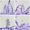
Light microscope images of linear glandular trichomes from phyllaries (a,d) and leave veins (b,c) in different developmental stages stained with Toluidine Blue O. (a) Early stage of a biosynthetically active LGT (between two nonglandular trichomes) showing strong vacuolization in the basal (B) and stalk cells (S), followed by five glandular cells rich in plasma and organelles and the apical cell starting with formation of a subcuticular cavity (black arrow). (b) Advanced stage of a biosynthetic activity LGT as indicated by progressed separation of the cuticle and extracellular storage of metabolites at the tip (black arrow). The apical cell had started degeneration and the wall partly collapsed. (c) Deposition of metabolites and cell degeneration (marked by stars) in the apical cell (A) and the adjacent glandular cell (G) while lower glandular cells still showed intact plastids (black arrowheads). The stalk cell showed thick lateral cell walls. (d) Senescent, degenerated trichome of the post-secretory stage with collapsed glandular cells but still vital stalk (S) and basal (B) cell in the epidermis (E). Scale bar = 50 µm.
Figure 3
Figure 3
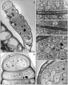
TEM images of a linear glandular trichome at the transition from the pre-secretory (a) to the secretory (b) stage. The overview (a) shows a young fully developed trichome anchored in the epidermis (EC) of an involucral bract at the sunflower capitulum. The trichome consisted of a basal cell (B), a stalk cell (S), 7 glandular cells and the apical cell (A). Vacuolization had just started in the basal and stalk cells, the apical cell did not yet show structures of compound accumulation or organ degradation and had not yet developed a subcuticular cavity. Chloroplasts (CP) in the glandular cells showed large electron-dense inclusions. (c) Cell wall between the apical cell (A) and uppermost glandular cell with plasmodesmata (white arrows). The apical cell contained many mitochondria (stars) and an extended rough endoplasmic reticulum (black arrows) close to the cell wall (CW). In the adjacent glandular cell, the nucleus (N) was visible. (d) Cell wall (CW) between two glandular cells from the middle of the trichome showing densely located plasmodesmata (white arrows) and mitochondria (stars). (b,e,f) Apical cell and glandular cells at the early secretory stage of development. The apical cell showed numerous mitochondria (stars), large amounts of rough endoplasmic reticulum (black arrows) and Golgi vesicles (black arrowheads). The cuticle started to separate (white arrowhead) from the cell wall to form a subcuticular cavity (SC). Scale bar: 10 µm (a)/5 µm (b)/1 µm (cf).
Figure 4
Figure 4
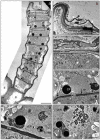
TEM images of a linear glandular trichome from a leaf vein in a late secretory stage. Frames in the overview (a) mark positions of the details (bf). (b) The apical cell (A) showed strong degeneration and was partly collapsed; the subcuticular cacity (SC) between the cell wall (CW) and the cuticle (white arrowheads) seemed to have collapsed through rupture of the cuticle. (c) The cytosol of the apical cell (A) lacked clearly defined organelles, whereas the adjacent glandular cell was still vital and contained an intact nucleus (N), numerous mitochondria (stars) and smooth (sER) endoplasmic reticulum. (d,e) Glandular cells in the upper and central part of the trichome showing plastids (CP) with electron-dense inclusions and plasmodesmata (black arrows) in the periclinal cell wall (CW). (f) Glandular cell in the lower trichome region baring crystals (PC; presumably of proteins) in the nucleus. The number and volume of vacuoles increased towards the trichome base. Scale bar: 10 µm (a)/1 µm (bf); (E) epidermis cell, (B) basal cell, (S) stalk cell.
Figure 5
Figure 5
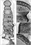
TEM images of a linear glandular trichome from a leaf vein in a late secretory stage. Frames in the photomerged overview (a,b) mark positions of the details (cf). (a) The three uppermost cells were filled with metabolites, the cellular compartmentation had collapsed and organelles had disintegrated. (c) The cell wall (CW) of the apical cell showed signs of loosening (white arrowheads), possibly indicating the formation of a subcuticular cavity. (d) The process of cellular degeneration progressed downwards from the third to the fourth cell (in which mitochondria (stars) and endoplasmic reticulum (ER) were still visible along with large amounts small vacuoles and vesicles (white arrowheads)). (e) Vital cells in the lower part of the trichome showed intact plastids (PL), mitochondria (stars) and endoplasmic reticulum (ER). (f) The stalk cell (S) and the basal cell (B) were characterized by large vacuoles (V) and numerous plasmodesmata (white arrows) in the thickened cell wall (CW). Scale bar: 10 µm (a,b)/1 µm (cf).

References

    1. Wagner G.J., Wang E., Shepherd R.W. New approaches for studying and exploiting an old protuberance, the plant trichome. Ann. Bot. 2004;93:3–11. doi: 10.1093/aob/mch011. - DOI - PMC - PubMed
    1. Werker E. Trichome diversity and development. Adv. Bot. Res. 2000;31:1–35. doi: 10.1016/s0065-2296(00)31005-9. - DOI
    1. Muravnik L.E. The structural peculiarities of the leaf glandular trichomes: A review. In: Ramawat K.G., Ekiert H.M., Goyal S., editors. Plant Cell and Tissue Differentiation and Secondary Metabolites, Reference Series in Phytochemistry. Springer Nature; Cham, Switzerland: 2020. pp. 1–34. - DOI
    1. Dai X., Wang G., Yang D.S., Tang Y., Broun P., Marks M.D., Sumner L.W., Dixon R.A., Zhao P.X. TrichOME: A comparative omics database for plant trichomes. Plant Physiol. 2010;152:44–54. doi: 10.1104/pp.109.145813. - DOI - PMC - PubMed
    1. Croteau R.B., Davis E.M., Ringer K.L., Wildung M.R. (−)-Menthol biosynthesis and molecular genetics. Naturwissenschaften. 2005;92:562–577. doi: 10.1007/s00114-005-0055-0. - DOI - PubMed

LinkOut - more resources