. 2021 Aug 20;10(8):1725.
doi: 10.3390/plants10081725.

In Vitro Evaluation of the Inhibitory Activity of Different Selenium Chemical Forms on the Growth of a Fusarium proliferatum Strain Isolated from Rice Seedlings

Affiliations

In Vitro Evaluation of the Inhibitory Activity of Different Selenium Chemical Forms on the Growth of a Fusarium proliferatum Strain Isolated from Rice Seedlings

Elisabetta Troni et al. Plants (Basel). .

Abstract

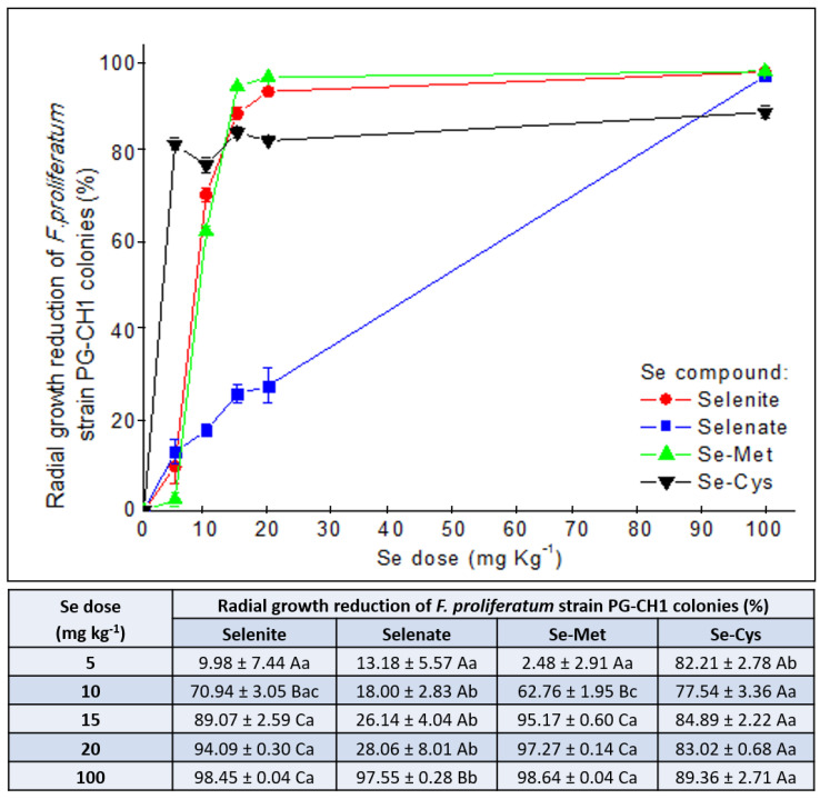
In this study, the in vitro effects of different Se concentrations (5, 10, 15, 20, and 100 mg kg-1) from different Se forms (sodium selenite, sodium selenate, selenomethionine, and selenocystine) on the development of a Fusarium proliferatum strain isolated from rice were investigated. A concentration-dependent effect was detected. Se reduced fungal growth starting from 10 mg kg-1 and increasing the concentration (15, 20, and 100 mg kg-1) enhanced the inhibitory effect. Se bioactivity was also chemical form dependent. Selenocystine was found to be the most effective at the lowest concentration (5 mg kg-1). Complete growth inhibition was observed at 20 mg kg-1 of Se from selenite, selenomethionine, and selenocystine. Se speciation analysis revealed that fungus was able to change the Se speciation when the lowest Se concentration was applied. Scanning Electron Microscopy showed an alteration of the fungal morphology induced by Se. Considering that the inorganic forms have a higher solubility in water and are cheaper than organic forms, 20 mg kg-1 of Se from selenite can be suggested as the best combination suitable to inhibit F. proliferatum strain. The addition of low concentrations of Se from selenite to conventional fungicides may be a promising alternative approach for the control of Fusarium species.

Keywords: Fusarium; bioactivity; fungi; inhibition; micronutrient; selenium.


Conflict of interest statement

The authors declare no conflict of interest.

Figures

Figure 1
Figure 1
Phylogenetic relationship of PG-CH1 strain (triangle) and members of the Fusarium fujikuroi species complex shown in a maximum likelihood dendrogram based on the Kimura 2-parameter model. The tree with the highest log likelihood (–1567.09) is shown. Bootstrap values are indicated above the branch nodes. A discrete Gamma distribution was used to model evolutionary rate differences among sites.
Figure 2
Figure 2
Effect of increasing selenium concentrations from different selenium forms on the colony development of Fusarium proliferatum strain PG–CH1 after 10 days of incubation at 22 ± 2 °C in the dark in comparison to the untreated control. Selenite (a), selenate (b), selenomethionine (c), selenocystine (d).
Figure 3
Figure 3
Effect of five selenium (Se) concentrations (5, 10, 15, 20, and 100 mg kg−1) from sodium selenite (selenite; red circles), sodium selenate (selenate; blue squares), selenomethionine (Se–Met; green up–triangles), and selenocystine (Se-Cys; black down-triangles) on Fusarium proliferatum strain PG-CH1 colony development. The inhibitory activity was measured after 10 days of incubation at 22 ± 2 °C in the dark and expressed as the mean (± standard error) of colony radial growth reduction (%) relative to the untreated control (0 mg kg−1 of Se), calculated according to the equation: (radial growthcontrol − radial growthtreatment): radial growthcontrol × 100. One-way ANOVA was used to determine statistically significant differences in radial growth of the tested strain. In the table, within a Se form (A–C) or a Se concentration (a–b), means with the same letter are not significantly different at p < 0.05 based on the Tukey Honestly Significant Difference multiple comparison test.
Figure 4
Figure 4
Selenium (Se) speciation data showing different Se chemical forms and their distribution after 10 days of incubation in the growth medium (potato dextrose agar) in the presence of Fusarium proliferatum strain PG-CH1 and following the application of increasing Se concentrations (5, 10, 15, 20, and 100 mg kg−1) as sodium selenite (selenite; a), sodium selenate (selenite; b), selenomethionine (Se-Met; c), and selenocystine (Se-Cys; d).
Figure 5
Figure 5
Scanning Electron Microscopy images showing Fusarium proliferatum strain PG-CH1 hyphae grown for 10 days on potato dextrose agar (PDA) not amended with selenium (Se) (labelled as control) (ac) and on PDA amended with 20 mg kg−1 of Se as sodium selenite (selenite; labelled as selenite 20) (df). The effect of 20 mg kg−1 of Se as selenite caused appreciable modifications in the morphology and density of F. proliferatum strain PG-CH1 hyphae (df) in comparison to the untreated control (ac).

References

    1. Steinbrenner H., Speckmann B., Klotz L.O. Selenoproteins: Antioxidant selenoenzymes and beyond. Arch. Biochem. Biophys. 2016;595:113–119. doi: 10.1016/j.abb.2015.06.024. - DOI - PubMed
    1. Roman M., Jitaru P., Barbante C. Selenium biochemistry and its role for human health. Metallomics. 2014;6:25–54. doi: 10.1039/C3MT00185G. - DOI - PubMed
    1. D’Amato R., Regni L., Falcinelli B., Mattioli S., Benincasa P., Dal Bosco A., Pacheco P., Proietti P., Troni E., Santi C., et al. Current knowledge on selenium biofortification to improve the nutraceutical profile of food: A comprehensive review. J. Agric. Food Chem. 2020;68:4075–4097. doi: 10.1021/acs.jafc.0c00172. - DOI - PMC - PubMed
    1. Ursini F., Maiorino M. Encyclopedia of Biological Chemistry. 2nd ed. Elsevier Inc.; Amsterdam, The Netherlands: 2013. Glutathione peroxidases; pp. 399–404.
    1. Gupta M., Gupta S. An overview of selenium uptake, metabolism, and toxicity in plants. Front Plant. Sci. 2017;7:2074. doi: 10.3389/fpls.2016.02074. - DOI - PMC - PubMed

LinkOut - more resources