. 2021 Aug 23;10(8):1740.
doi: 10.3390/plants10081740.

Induction of Conjugation and Zygospore Cell Wall Characteristics in the Alpine Spirogyra mirabilis (Zygnematophyceae, Charophyta): Advantage under Climate Change Scenarios?

Affiliations

Induction of Conjugation and Zygospore Cell Wall Characteristics in the Alpine Spirogyra mirabilis (Zygnematophyceae, Charophyta): Advantage under Climate Change Scenarios?

Charlotte Permann et al. Plants (Basel). .

Abstract

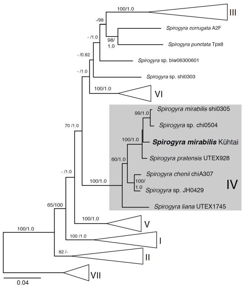
Extreme environments, such as alpine habitats at high elevation, are increasingly exposed to man-made climate change. Zygnematophyceae thriving in these regions possess a special means of sexual reproduction, termed conjugation, leading to the formation of resistant zygospores. A field sample of Spirogyra with numerous conjugating stages was isolated and characterized by molecular phylogeny. We successfully induced sexual reproduction under laboratory conditions by a transfer to artificial pond water and increasing the light intensity to 184 µmol photons m-2 s-1. This, however was only possible in early spring, suggesting that the isolated cultures had an internal rhythm. The reproductive morphology was characterized by light- and transmission electron microscopy, and the latter allowed the detection of distinctly oriented microfibrils in the exo- and endospore, and an electron-dense mesospore. Glycan microarray profiling showed that Spirogyra cell walls are rich in major pectic and hemicellulosic polysaccharides, and immuno-fluorescence allowed the detection of arabinogalactan proteins (AGPs) and xyloglucan in the zygospore cell walls. Confocal RAMAN spectroscopy detected complex aromatic compounds, similar in their spectral signature to that of Lycopodium spores. These data support the idea that sexual reproduction in Zygnematophyceae, the sister lineage to land plants, might have played an important role in the process of terrestrialization.

Keywords: Spirogyra; alpine region; cell wall; conjugation; sexual reproduction; streptophyte; zygospore.


Conflict of interest statement

The authors declare no conflict of interest.

Figures

Figure 1
Figure 1
Habitat of isolated Spirogyra mirabilis: (a) sampling site with zygnematophycean green algal mat, (b) isolated unialgal culture, (c) detailed view of vegetative filament. Scale bars (b) 1 mm, (c) 20 µm.
Figure 2
Figure 2
Conjugation and zygospores of Spirogyra mirabilis: (a) lateral conjugation, (b) scalariform conjugation, (c) zygospore formed by lateral conjugation, (d) zygospores formed by scalariform conjugation, (e) polymorphic zygospores, (f) germinating zygospore, (g) vegetative filament, (h) vegetative filament stained with Indian ink, (i) zygospore stained with Indian ink, (j) zygospores with chloroplast with (k) corresponding chlorophyll autofluorescence image, (l) semi-thin section with (m) corresponding toluidine staining. Scale bars 20 µm.
Figure 3
Figure 3
Summary of the maximum likelihood analysis of rbcL and atpB sequences from Spirogyra mirabilis (Kühtai) in the context of related and sequenced Spirogyra strains in clade IV, and for which the full tree is presented in Supplementary Figure S2. The tree is oriented, and the major clades are labeled, after Stancheva et al. [27] and Takano et al. [38]. Node support values correspond to ML bootstrap/BPP values. Scale bar indicates the expected number of substitutions/sites.
Figure 4
Figure 4
Transmission electron micrographs of zygospores of Spirogyra mirabilis: (a) gametangia with zygospore and conjugation tube, (b) overview of mature zygospore, (c) mature zygospore with lipid accumulation, (d) mitochondria and lipid bodies in the cell lumen, (e) detailed view of the chloroplast lobes with plastoglobuli, (f) nucleus with nucleolus, (g) detailed view of the zygospore wall of a presumably young zygospore with visible exospore structure, (h) mature zygospore wall, (i) mature zygospore with distinct orientation of the microfibrils of the endospore. Abbreviations: Chl—chloroplast, CT—conjugation tube, CW—cell wall of the gametangium, En—endospore, Ex—exospore, L—lipid bodies, Me—mesospore, M—mitochondrion, N—nucleus, Nu—nucleolus, PG—plastoglobuli. Scale bars (a) 5 µm, (b,c) 2500 nm, (d,f,i) 1 µm, (e,g,h) 500 nm.
Figure 5
Figure 5
Determining cell wall epitopes in Spirogyra mirabilis using carbohydrate microarray profiling. A total of >40 cell wall probes were incubated with CDTA and NaOH extracts sequentially prepared from Spirogyra AIR. The heatmap color intensity shows the strength of the probe binding. The strongest signal was assigned a value of 100 and the cut-off signal was set to 5. Probe codes are in bold and bound epitopes in brackets. Abbreviations: Ara—arabinose, DE—degree of esterification, Fuc—fucose, Gal—galactose, GalA—galacturonic acid, GalNAc—N-acetylgalactosamin, GlcA—glucuronic acid, HG—homogalacturonan, RGI—rhamnogalacturonan I, Rha—rhamnose, AGPs—arabinogalactan proteins. Oligosaccharide nomenclature for xyloglucan probes, see Fry et al. [50].
Figure 6
Figure 6
Immuno-localization of certain cell wall components in Spirogyra mirabilis. (a, b) JIM13 (AGP) and (c) LM15 (xyloglucan) of semi-thin sections of Spirogyra mirabilis, (a) zygospore in gametangium, (b) conjugating cells, (c) bilayer arrangement in zygospores wall. Scale bars 20 µm.
Figure 7
Figure 7
Raman imaging of Spirogyra mirabilis zygospores reveals aromatic compounds in the cell wall. (a) Brightfield image of the investigated spore (white rectangle). (b) Confocal Raman images with a depth profiling of 9 μm, measured as nine layers in a stack for every micrometer in the z-direction, up to down. (c) Raman fluorescence image of the middle layer of the spore. (d) Aromatic compounds impregnate the cell wall as visualized by integrating the peak at 1604 cm−1. (e) Magnification of the cell wall shows high intensity of aromatic compounds in the zygospore cell wall, but no clear layering. (f) The extracted average spectrum from the Spirogyra cell wall (red line) shows clear aromatic bands around 1600 cm−1. Fitting of reference Raman spectra by a linear combination using the Orthogonal Matching Pursuit Method confirmed the aromatic nature and placed the Lycopodium spore cell wall spectrum on top with the highest contribution (similarity) (Supplementary Figure S4).

Similar articles

Cited by

References

    1. Wodniok S., Brinkmann H., Glöckner G., Heidel A.J., Philippe H., Melkonian M., Becker B. Origin of land plants: Do conjugating green algae hold the key? BMC Evol Biol. 2011;11:104. doi: 10.1186/1471-2148-11-104. - DOI - PMC - PubMed
    1. de Vries J., Archibald J.M. Plant evolution: Landmarks on the path to terrestrial life. New Phytol. 2018;217:1428–1434. doi: 10.1111/nph.14975. - DOI - PubMed
    1. Leebens-Mack J.H., Barker M.S., Carpenter E.J., Deyholos M.K., Gitzendanner M.A., Graham S.W., Grosse I., Li Z., Melkonian M., Mirarab S., et al. One thousand plant transcriptomes and the phylogenomics of green plants. Nature. 2019;574:679–685. doi: 10.1038/s41586-019-1693-2. - DOI - PMC - PubMed
    1. Cheng S., Xian W., Fu Y., Marin B., Keller J., Wu T., Sun W., Li X., Xu Y., Zhang Y., et al. Genomes of subaerial Zygnematophyceae provide insights into land plant evolution. Cell. 2019;179:1057–1067.e14. doi: 10.1016/j.cell.2019.10.019. - DOI - PubMed
    1. Jiao C., Sørensen I., Sun X., Sun H., Behar H., Alseekh S., Philippe G., Lopez K.P., Sun L., Reed R., et al. The Penium margaritaceum genome: Hallmarks of the origins of land plants. Cell. 2020;181:1097–1111. doi: 10.1016/j.cell.2020.04.019. - DOI - PubMed

LinkOut - more resources